

Vision Starts With Light and Ends With Perception
Imagine standing on a sunny beach and watching waves roll toward the shore. Your eyes transform light bouncing off those waves into electrical signals that your brain interprets as colour, shape and movement. This complex process happens so quickly and seamlessly that we rarely think about it. Understanding how light becomes vision can help us appreciate the fragility of our eyes and encourage us to take good care of them.
This guide explains how the human eye turns light into sight. The information has been simplified to be readable at a seventh‑grade level while still providing enough depth for curious readers. We’ll explore the anatomy of the eye, the journey of light through the eye’s structures, the roles of rods and cones, how signals travel through the optic nerve to the brain and how colour vision works. Along the way you’ll find high‑quality external links for more information and suggestions for internal links to related topics like colour blindness and macular degeneration.
A Look at Eye Anatomy
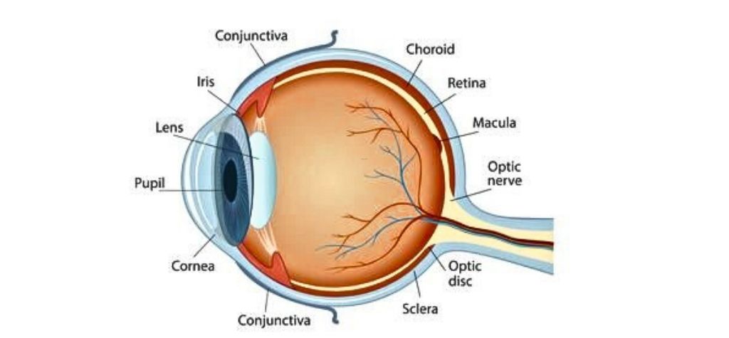
Before diving into the path of light, it helps to know the main parts of the eye. Four structures work together to form an image: the cornea, pupil, lens and retina. The cornea is the clear, dome‑shaped front layer. It bends incoming light to begin focusing it. Behind the cornea is the pupil, the circular opening that lets light into the eye. The coloured iris controls how wide or narrow the pupil becomes. In bright conditions the iris squeezes the pupil to protect the retina from too much light; in dim conditions it expands the pupil to let in more light.
Next is the lens, a transparent structure just behind the pupil. Tiny muscles called ciliary muscles change the lens’s shape so that light rays from near and far objects focus precisely on the back of the eye. This focusing process is called accommodation. Finally, at the back of the eye lies the retina, a thin layer of nerve tissue containing millions of photoreceptor cells. We’ll look more closely at the retina later because it’s where light is converted to electricity. The space between the lens and retina is filled with the vitreous body, a gel that helps the eye keep its shape.
Protective and Support Structures
Several supportive structures protect and nourish the eye:
- Sclera: The tough white outer layer that gives the eye its shape.
- Choroid: A layer of blood vessels between the sclera and retina that provides oxygen and nutrients to the eye.
- Aqueous humor: A clear fluid in front of the lens that nourishes the cornea and lens.
- Vitreous humor: The gel‑like substance filling the main cavity of the eye, maintaining its form.
- Conjunctiva: A thin membrane that covers the white of the eye and inside of the eyelids.
- Eyelids and eyelashes: These keep dust and debris out and spread lubricating tears across the surface.
The macula is a small region near the centre of the retina. Within it is the fovea, a tiny pit containing only cones. This area provides the sharpest vision. Damage to the macula leads to central vision loss, a condition called macular degeneration.
The Path of Light Through the Eye
Step 1: Light Enters Through the Cornea and Pupil
Light waves enter the eye when they pass through the cornea. The cornea bends these rays and directs them through the pupil. The iris adjusts the pupil size to control how much light gets in. In bright sunshine the pupils constrict; in a dark room they dilate.
Fun fact: The term pupil comes from the Latin word for “doll” because ancient people noticed tiny reflections of themselves in another person’s eyes.
Step 2: The Lens Focuses Light on the Retina
After passing through the pupil, light hits the lens. Working with the cornea, the lens fine‑tunes the focus so the incoming image lands sharply on the retina. Ciliary muscles change the lens’s curvature—more curved to focus on close objects and flatter for distant objects.
Step 3: Photons Reach the Retina
When light reaches the retina, it passes through several layers of transparent cells before hitting photoreceptors at the back of the retina. The two types of photoreceptors—rods and cones—contain light‑sensitive pigments that absorb photons and trigger chemical reactions. This is the beginning of phototransduction, the process that converts light into electrical signals.
Photoreceptors: Rods and Cones
Why Two Types of Photoreceptors?
The retina’s two kinds of sensory cells—rods and cones—serve different purposes. Rods help us see shapes and movement in dim light. Cones allow us to see fine detail and colour under bright light. Together they enable vision across a wide range of lighting conditions.
Rods
- There are over 100 million rods in each human eye.
- Rods are extremely sensitive to light; only a few photons can activate them.
- They do not detect colour, so images appear in shades of grey at night or in dark environments.
- Rods are concentrated toward the peripheral retina. This is why your peripheral vision works better in low light.
Rods contain a pigment called rhodopsin, which changes shape when it absorbs light. This shape change triggers a cascade of chemical events that ultimately generate an electrical signal. When rhodopsin absorbs a photon, it splits into a protein (opsin) and a vitamin A derivative called retinal. Vitamin A is therefore vital for rod function, which is why severe vitamin A deficiency causes night blindness.
Cones
- Humans have about 6 million cone cells, far fewer than rod.
- Cones require bright light to function; they shut down at night, which is why colours fade in dim conditions.
- Cones are densest in the fovea—the centre of your visual field—and are responsible for sharp, detailed vision.
- Each cone is sensitive to either short (blue), medium (green) or long (red) wavelengths of light. By comparing signals from these three cone types, the brain perceives a full spectrum of colours.
Cones contain photopsins, pigments tuned to specific wavelengths. When light hits a photopsin, it changes shape and activates a similar chemical cascade as rods, but the details differ. Cones respond faster than rods and recover more quickly between flashes of light.
Phototransduction: Converting Light to Electricity
When light strikes rhodopsin or photopsin molecules in the outer segments of rods and cones, the molecules change shape and set off a chain reaction that opens and closes ion channels in the cell membrane. The resulting change in electric charge across the photoreceptor’s membrane is the first step in converting photons into neural signals. The signal travels from the photoreceptor’s outer segment to its inner end and then passes to bipolar cells and ganglion cells. Ganglion cell axons converge to form the optic nerve.
From Retina to Brain: The Visual Pathway
Optic Nerve and Beyond
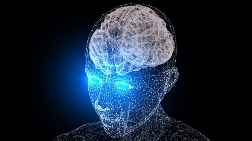
The optic nerve carries signals from each retina to the brain. Because the nerves cross at the optic chiasm, each half of the brain processes information from the opposite visual field. Signals then travel through relay stations in the thalamus to the visual cortex in the occipital lobe, where basic shapes, edges and colours are decoded. Higher processing areas in the brain integrate these signals, combine information from both eyes and compare them with memory and expectations to create the final image we perceive.
Parallel Pathways
Visual information travels along multiple pathways. One pathway carries information about shape and colour via the parvocellular system, while another carries information about motion and contrast via the magnocellular system. Separate pathways enable the brain to process different aspects of a scene simultaneously, giving us a rich, detailed picture of the world. Damage to specific pathways can cause deficits such as colour blindness or motion blindness.
How Colour Vision Works
Colour vision arises because cones come in three varieties—S‑cones, M‑cones and L‑cones—each sensitive to different wavelength ranges. S‑cones peak at about 426 nm (blue), M‑cones at 530 nm (green) and L‑cones at 555 nm (red). Your brain determines a colour by comparing the relative activation of these cone types. For example, yellow light stimulates red and green cones but not blue cones, so the brain perceives yellow.
Colour Opponency and Brain Processing
After initial phototransduction, signals from cones are organized in opponent channels. One channel contrasts red versus green, another contrasts blue versus yellow and a third contrasts light versus dark. These opponent processes begin in the retina and continue in the lateral geniculate nucleus (LGN) and visual cortex. Opponent coding helps explain why certain colour combinations (such as “reddish‑green”) don’t exist and why afterimages occur when you stare at a colour for a long time.
Colour Vision Deficiencies
People with colour vision deficiencies lack one or more cone types or have altered photopigments. Red‑green colour blindness, the most common type, results from problems with L‑ or M‑cones. It is inherited in an X‑linked pattern, affecting more men than women. Blue‑yellow colour blindness is rarer and involves S‑cone defects. Complete colour blindness (achromatopsia) occurs when all cones are absent or non‑functional. Many types of colour vision deficiency can be detected with simple screening tests such as Ishihara plates or Farnsworth panels.
How We Perceive Depth and Motion
The eyes are positioned slightly apart, so each retina receives a slightly different image. The brain compares these images in the visual cortex to extract depth information, a process called stereopsis. Other depth cues include motion parallax (closer objects move faster across our visual field), relative size, shading and perspective.
Motion perception relies on specialised cells in the retina and brain that respond to moving objects. The magnocellular pathway is particularly important for motion detection. When these pathways malfunction, as in certain neurological diseases, people may struggle to perceive movement smoothly.
Common Eye Disorders Affecting Vision
Refractive Errors
Not all eyes focus light perfectly. Myopia (nearsightedness) occurs when the eye is too long or the cornea too curved, causing light to focus in front of the retina, making distant objects blurry. Hyperopia (farsightedness) happens when the eye is too short or the cornea too flat, causing light to focus behind the retina. Astigmatism arises when the cornea has an irregular shape, leading to distorted images. These errors can often be corrected with glasses, contact lenses or laser surgery.
Cataracts
A cataract is a clouding of the lens that blocks or scatters light. It usually develops slowly with age. Surgery can replace the cloudy lens with a clear artificial one.
Glaucoma
Glaucoma is a group of diseases in which elevated pressure inside the eye damages the optic nerve. Without treatment, it can lead to peripheral vision loss and eventually blindness. Regular eye exams are essential for early detection.
Macular Degeneration
Age‑related macular degeneration (AMD) damages the macula and fovea, impairing central vision. People with AMD may see a dark spot in the centre of their visual field or notice that straight lines look wavy. There are two forms—dry AMD, which progresses slowly, and wet AMD, which can cause rapid vision loss. Healthy lifestyle choices and prompt treatment can slow the disease.
Diabetic Retinopathy
High blood sugar damages blood vessels in the retina, causing leakage or abnormal growth of new vessels. Over time, this can lead to vision loss. Strict control of blood sugar, blood pressure and cholesterol helps prevent or slow diabetic retinopathy.
Retinal Detachment
Retinal detachment occurs when the retina separates from the underlying tissue. Symptoms include flashing lights, a sudden increase in floaters or a shadow across your vision. It requires urgent medical attention to prevent permanent vision loss.
Colour Vision Disorders
As discussed earlier, colour vision deficiencies result from missing or malfunctioning cone cells. People with colour blindness may have difficulty distinguishing certain colours but usually have normal visual acuity. Special lenses and apps can help in some cases.
Taking Care of Your Eyes
Maintaining good eye health can help preserve vision for decades. Here are practical tips:
- Get regular eye exams: Even if you see well, comprehensive exams can detect problems like glaucoma or diabetic retinopathy before symptoms appear.
- Eat a balanced diet: Foods rich in vitamin A (carrots, sweet potatoes), omega‑3 fatty acids (fish) and antioxidants (leafy greens) support eye health.
- Protect eyes from UV light: Wear sunglasses that block 99–100 % of UV‑A and UV‑B rays.
- Manage chronic conditions: Control blood sugar, blood pressure and cholesterol to reduce the risk of diabetic retinopathy and hypertension‑related eye diseases.
- Take breaks from screens: Follow the 20‑20‑20 rule—every 20 minutes, look at something 20 feet away for 20 seconds to reduce digital eye strain.
- Don’t smoke: Smoking increases the risk of cataracts, macular degeneration and other eye diseases.
Conclusion: Seeing Is a Team Effort
Vision is not just about the eye itself. It’s a partnership between the eye and the brain. Light passes through the cornea and lens, the retina’s rods and cones convert photons to electrical signals, the optic nerve carries these signals and the brain assembles them into a coherent picture. Each component plays a vital role. The eye’s delicate structures are susceptible to disease and injury, but many problems can be prevented or treated with early detection and healthy habits.
Understanding how the eye actually sees helps us appreciate the complexity of our visual system. It also underscores why regular check‑ups, proper nutrition and protective measures are essential for maintaining good vision throughout life.

")

