
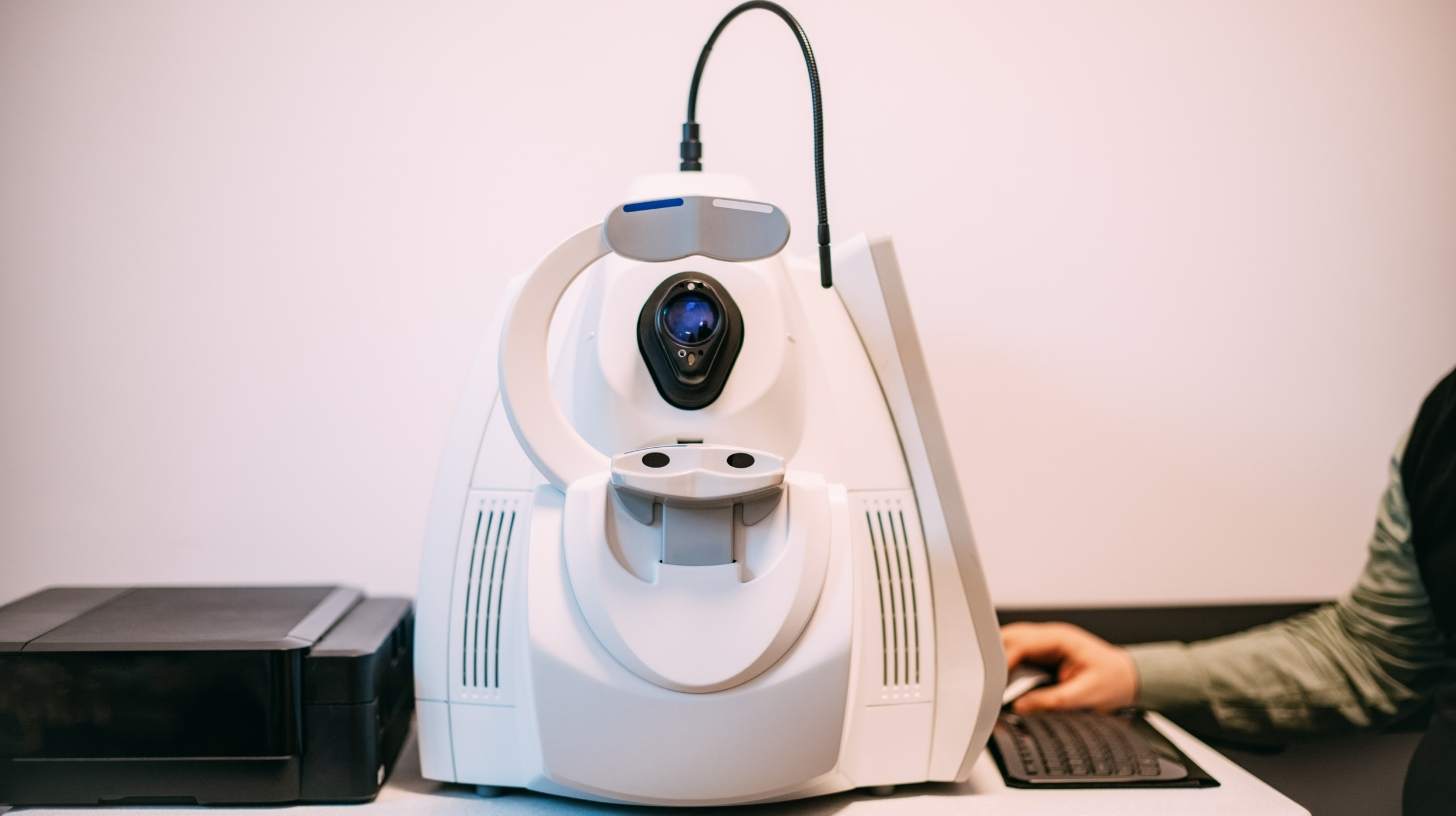
Has your eye doctor ordered an OCT eye scan? The name sounds technical, but the test itself is usually quick and simple.
You will sit at a camera-like machine, rest your chin, and look at a target. The device scans one eye at a time using light. Nothing usually touches your eye, and the scan should not hurt.
The image capture often takes only a few minutes. Your full visit may be longer if you need dilation, other tests, or a discussion with your doctor.
Here are seven things you can expect, from preparation through results.
What is an OCT eye scan?

OCT stands for optical coherence tomography. It is a noninvasive imaging test that creates detailed cross-section pictures of eye tissue.
Most eye OCT scans examine the retina and optic nerve. The retina is the light-sensitive tissue lining the back of the eye. The optic nerve carries visual signals from the eye to the brain.
An OCT eye scan uses reflected light to create layered pictures of the retina or optic nerve. It works somewhat like an optical ultrasound, but it uses light instead of sound.
The test can show separate retinal layers and measure their thickness. It may reveal swelling, thinning, fluid, or changes in shape. This plain-language explanation of optical coherence tomography describes the structures doctors can examine.
Some machines also scan the cornea and drainage angle at the front of the eye. The exact test depends on why you were referred and which equipment the clinic uses.
Why your eye doctor may recommend an OCT test
An OCT scan provides information that an ordinary vision chart cannot. It helps doctors examine tissue beneath the visible surface and compare small changes over time.
Your optometrist or ophthalmologist may order a scan to investigate or monitor:
- Age-related macular degeneration
- Diabetic retinal changes or macular swelling
- Glaucoma or possible optic nerve damage
- A macular hole or epiretinal membrane
- Fluid beneath or within the retina
- Retinal blood-vessel problems
- Unexplained changes in central vision
- Certain corneal or drainage-angle conditions
Having an OCT test does not mean you have one of these conditions. The doctor may want a baseline image for future comparison. A baseline is a record of how your eyes look at one point in time.
OCT may also be repeated during treatment or routine monitoring. A series of good-quality scans can be more useful than one isolated image.
7 things to expect during an OCT eye scan

Clinic routines differ, but most appointments follow the same basic pattern. Ask the office about any special instructions before arriving.
1. You may have a short check before the scan
The technician may confirm your name, symptoms, medical history, and reason for testing. Bring your current glasses and a list of medicines if the clinic requested them.
Tell the staff about recent eye surgery or difficulty sitting at a machine. Mention if you have trouble keeping your head still or focusing on a target. Small adjustments may make the scan easier.
You will probably remove your glasses before the image is taken. Whether contact lenses must come out depends on the scan and clinic procedure. Follow the staff’s instructions rather than removing them automatically.
No fasting, injections, or blood test is normally required for a standard OCT scan. Keep using prescribed eye medicines unless your clinician gave different instructions.
2. Your pupils may—or may not—be dilated
Many OCT scans can be completed through an undilated pupil. However, dilation may improve the view when pupils are small or an image is difficult to capture.
Your doctor may also want a dilated retinal examination during the same visit. Dilation uses eye drops to enlarge the pupils. This lets more light enter and gives a wider view of the back of the eye.
The drops can cause light sensitivity and blurry near vision for several hours. Effects vary by person and by the drops used. Bring sunglasses if the clinic says dilation is possible.
Ask in advance whether you may need someone to drive you. Some people feel comfortable driving after dilation, while others do not. Do not drive if your vision feels unsafe or your clinician advises against it.
3. You will sit at a camera-like machine
The OCT unit usually sits on a table. You will place your chin on a support and rest your forehead against a band. These supports help keep your head lined up.
The technician adjusts the machine to your height and eye position. Tell them if the chin rest, chair, or forehead band feels uncomfortable. They can often reposition it.
The camera does not need to press against the eye for a standard scan. You will remain awake and seated. No anesthetic drops are usually needed.
Wheelchair users and people with limited neck movement may need a different setup. Let the clinic know before the visit so it can plan enough time or suitable equipment.
4. You will look at a target while each eye is scanned
The technician will ask you to look at a small light, dot, picture, or cross. This fixation target keeps the desired part of the retina centered.
One eye is scanned while the other remains open or is temporarily covered. Try to look steadily at the target without following other lights inside the machine.
You may notice a bright line or brief flash. You might also hear the technician give simple directions. The device collects many light reflections and builds them into a layered image.
A typical scan is fast. The usual OCT scanning experience involves scanning one eye at a time while the patient stays still for a minute or two.
If central vision is poor, the target may be hard to see. Tell the technician rather than guessing where to look. They may use an outside target or guide your gaze in another way.
5. The test should be painless and noncontact
OCT uses light, not X-rays. It does not expose you to ionizing radiation. Nothing is injected for a routine structural OCT scan.
The light may seem bright, especially after dilation. That can feel uncomfortable for a moment, but the scan should not be painful. Tell the technician if you feel pain or become unwell.
Standard OCT is also different from fluorescein angiography. Angiography may use an injected dye to photograph blood flow. OCT angiography, available on some machines, can map movement in blood vessels without dye.
The type of scan ordered should be explained if you are unsure. Do not assume that every test called “retinal imaging” uses the same method.
6. Blinking or movement may lead to a repeat scan
You can blink normally while the technician prepares the machine. They may then ask you to hold your eyes open for a few seconds during capture.
A blink, eye movement, or head movement can leave a gap or distortion in the image. Dryness can also reduce picture quality. The technician may repeat a section to get a clearer result.
A repeat does not mean something is wrong with your eye. It often means the first image was not centered or clear enough. Several scans may also be taken using different patterns.
Clouding in the eye can make OCT imaging harder. A dense cataract, corneal scar, or bleeding inside the eye may block some light. Very small pupils and difficulty fixing on the target can also limit quality.
High-quality three-dimensional images of retinal layers and the optic nerve depend on a clear light path and steady positioning.
7. Your doctor will interpret maps, measurements, and images
OCT software displays cross-sections that may resemble colored layers or a landscape. It can also create thickness maps, graphs, and comparisons with a reference database.
Colors on an OCT report are not a diagnosis by themselves. Red does not always mean disease, and green does not always guarantee a healthy eye. Age, eye length, prescription, scan quality, and software boundaries can affect the display.
The clinician reviews the actual scan, not just its color code. They combine it with your symptoms, eye pressure, retinal examination, photographs, or visual-field testing when appropriate.
Previous images are especially helpful. Changes over time may matter more than a single measurement. Ask whether the scan is a baseline and when, if ever, it should be repeated.
What the OCT scan may show
Different scan programs answer different questions. A macular OCT focuses on the central retina. The macula supports detailed vision used for reading and recognizing faces.
The scan may show retinal swelling, a pocket of fluid, thinning, a hole, or a membrane pulling on the surface. These findings can have several causes and require clinical interpretation.
A glaucoma OCT often measures the retinal nerve fiber layer and other structures near the optic nerve. These are groups of nerve fibers that carry visual information. Thinning may support concern about glaucoma, but OCT alone does not prove the diagnosis.
Glaucoma assessment may also involve eye-pressure measurement, examination of the drainage angle, optic nerve evaluation, and visual-field testing. Readers can learn more about why glaucoma may develop before obvious symptoms.
Some scans examine the front of the eye. An anterior-segment OCT can measure parts of the cornea or show the drainage angle. It does not use the same scan pattern as a retinal OCT.
How long does an OCT appointment take?
The image capture may take only a few minutes. Allow more time for registration, positioning, dilation, repeat images, and other eye tests.
A clinic may perform OCT during a full eye examination. Another clinic may schedule imaging first and have a doctor review it later. Ask whether you will receive an explanation on the same day.
If dilation is needed, the drops may take about 15 to 30 minutes to work. Clinic flow and individual response can change that estimate.
Plan extra time when the scan is being done for the first time. Old scans may also need to be imported so the doctor can compare them correctly.
What happens after an OCT eye scan?
There is usually no recovery period after an undilated scan. You can return to normal activities unless the clinic gives other instructions.
If your pupils were dilated, near vision and light sensitivity may continue for several hours. Sunglasses can improve comfort outdoors. Avoid driving or hazardous work until you can see safely.
The technician may not be allowed to explain the medical meaning of the images. That does not signal a bad result. Image capture and clinical interpretation are different roles.
Your eye doctor may say that the scan looks stable, request another test, arrange monitoring, or discuss treatment. An abnormal-looking area sometimes turns out to be an imaging artifact. An artifact is a false or misleading feature caused by movement, poor focus, or software error.
The role of OCT in clinical review and treatment decisions shows why the image must be reviewed in the context of the full eye examination.
What OCT cannot tell your doctor by itself
OCT is powerful, but it is not a complete eye exam. It does not test every part of vision or every eye structure.
The scan does not replace:
- A visual acuity test to measure how clearly you see
- Refraction to determine a glasses prescription
- Eye-pressure measurement
- A slit-lamp examination of the front of the eye
- A full retinal examination when one is needed
- Visual-field testing for side vision
- A discussion of symptoms and medical history
Automated analysis can also make errors. The software may place a boundary on the wrong retinal layer. Unusual eye anatomy may not match the machine’s reference database.
Artificial intelligence can help flag patterns, but a professional still needs to review the scan. This fits with the broader limits explained in why imaging and AI cannot replace a complete eye examination.
How to prepare for your retinal scan
Most people need little preparation. Confirm the appointment time and ask whether dilation is planned.
It can help to:
- Bring glasses, sunglasses, and insurance information.
- Bring a medicine list if requested.
- Tell the clinic about mobility or fixation difficulties.
- Ask whether contact lenses should be removed.
- Arrange transport if dilation may make driving difficult.
- Bring previous eye records if the clinic does not already have them.
Do not stop prescribed medicines without medical instructions. Food and drink are usually allowed before a standard OCT test.
When symptoms need attention before the scheduled scan
An OCT appointment may be arranged for routine monitoring. However, some symptoms should not wait for a future appointment.
Seek urgent eye care for a sudden major loss of vision, a dark curtain or shadow, many new floaters, repeated flashes, or severe eye pain with redness. Sudden distortion or a new blank spot in central vision also deserves prompt assessment.
These symptoms do not identify one condition. They may signal problems that need a same-day examination. Call an eye clinic or emergency service for local guidance.
The practical takeaway
An OCT eye scan is usually quick, painless, and noncontact. Expect to rest your chin, look at a target, and have each eye scanned separately. Dilation is sometimes needed, but not for every scan.
The most important step happens after the pictures are taken. A trained clinician must interpret the layers, measurements, and image quality alongside the rest of your examination.
Before leaving, ask three questions: What part of my eye was scanned? Is this a baseline or a comparison? When will I receive the results?
Knowing those answers turns an unfamiliar test into a useful part of your eye-care plan.
Frequently asked questions
Does an OCT eye scan hurt?
No. A standard OCT eye scan is noncontact and should not hurt. You rest your head against supports and look into the machine. The light may seem bright, especially after dilation, but it lasts briefly. Tell the technician if you experience pain, feel unwell, or need a break.
Do I need eye drops before an OCT scan?
Not always. Many scans can be taken without drops. Dilation may be used when your pupils are small, image quality is poor, or your doctor also needs a wider retinal examination. Ask the clinic beforehand because dilation can blur near vision and increase light sensitivity for several hours.
How long does an OCT test take?
The scan itself often takes only a few minutes, with each eye captured separately. Your visit may take longer because of registration, positioning, dilation, repeat images, or other tests. If the clinic is reviewing several eye conditions or comparing old scans, allow additional time for discussion.
Can I drive after optical coherence tomography?
An undilated OCT scan does not usually affect driving. Dilation can make vision blurry and light sensitive, so driving may be unsafe for some people. Ask whether dilation is planned and arrange transport if needed. Never drive until your vision feels clear enough and your clinician’s instructions allow it.
Can an OCT scan diagnose glaucoma?
OCT can show thinning around the optic nerve and help monitor change. However, it does not diagnose glaucoma by itself. Doctors also consider eye pressure, optic nerve appearance, drainage-angle findings, visual fields, family history, and other risk factors. An unusual OCT measurement needs interpretation within a complete evaluation.
What is the difference between OCT and a retinal photograph?
A retinal photograph records a two-dimensional view of the retina’s surface. OCT creates cross-section images showing separate tissue layers and their thickness. The tests provide different information and may be used together. Neither test replaces every part of a comprehensive eye examination.
Why did the technician repeat my OCT scan?
A blink, eye movement, poor focus, small pupil, or positioning problem can reduce image quality. The technician may repeat the scan to center it or remove gaps. A repeat usually reflects a technical issue, not proof of disease. Some appointments also require several scan patterns for different structures.