

Have you ever stared at a clock and realized you can’t see the hands even though the numbers are clear? Or perhaps you noticed that straight lines suddenly look wavy, like ripples on a pond. These strange visual experiences can be the first signs of macular degeneration, an eye disease that damages the part of the retina responsible for sharp central vision. When the macula deteriorates, people may still have peripheral (side) vision but lose the ability to read, drive or recognize faces. Age‑related macular degeneration (AMD) is the most common form of macular degeneration and a leading cause of vision loss in older adults. Around 20 million U.S. adults are living with macular degeneration today, and projections suggest that 288 million people worldwide could have the condition by 2040.
This comprehensive guide explains how macular degeneration affects the eyes, explores different types and stages, discusses causes and risk factors, outlines current and emerging treatments, and offers practical tips for prevention and daily living. The content is written in a conversational style with straightforward language to ensure a seventh‑grade reading level. Whether you or a loved one has been diagnosed with AMD or you simply want to safeguard your vision, this article will provide the information you need to make informed choices.
Understanding Eye Anatomy: The Retina and the Macula
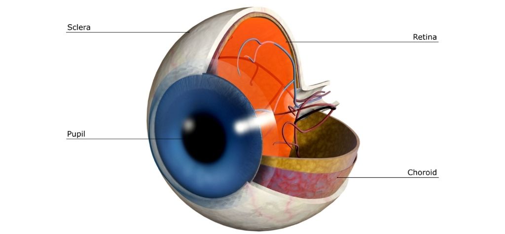
To appreciate how macular degeneration develops, it helps to understand basic eye anatomy. At the back of your eye is a light‑sensitive tissue called the retina. When light enters the eye, the retina converts it into electrical signals that travel to the brain via the optic nerve. Within the retina lies a small but critical region called the macula. The macula contains a high concentration of cone photoreceptors, which are cells responsible for perceiving fine details and color. When the macula functions properly, you can read small print, thread a needle and recognize faces.
In macular degeneration, the cells of the macula become damaged or die. As a result, central vision deteriorates, leaving a blind or blurry spot at the center of one’s visual field. Peripheral vision remains intact, so people do not go completely blind, but losing central vision significantly affects day‑to‑day tasks such as reading and driving. Many people with AMD feel like they are seeing through a blurry or dark hole in the middle of their vision.
Types of Macular Degeneration
Macular degeneration is not a single disease but a group of disorders that cause macular damage. Understanding the differences between these types can help you and your healthcare provider tailor an appropriate management plan.
Dry (Atrophic) Age‑Related Macular Degeneration
Dry AMD, also known as atrophic AMD, is the most common form and accounts for about 80–90 percent of cases. It develops when the macula gradually thins with age, and tiny yellow deposits called drusen accumulate under the retina. In early stages, drusen are small and few, and vision remains normal. As drusen enlarge and multiply, they interfere with the function of retinal cells, leading to gradual central vision loss.
Dry AMD progresses slowly and typically has three stages:
- Early dry AMD – Drusen are present, but there are no noticeable symptoms. Regular eye exams may detect subtle changes before vision is affected.
- Intermediate dry AMD – Drusen grow larger or more numerous, and pigment changes may occur in the retina. Some people notice mild blurriness or difficulty seeing in low light.
- Late dry AMD (Geographic Atrophy) – Sections of macular tissue die (called geographic atrophy), causing blind spots in the central vision. Colors may appear less vivid. Late dry AMD can progress to wet AMD in some cases.
Wet (Neovascular) Age‑Related Macular Degeneration
Wet AMD, also known as neovascular AMD or exudative AMD, is less common, affecting around 10–20 percent of people with AMD. However, it is responsible for the majority of severe vision loss. Wet AMD occurs when abnormal blood vessels grow under the retina and the macula. These vessels are fragile and leak blood or fluid, leading to swelling, scarring and rapid vision loss. Symptoms often appear suddenly: straight lines look wavy, dark spots appear in the center of vision, and colors seem faded or washed out. Without prompt treatment, wet AMD can cause significant loss of central vision.
Other Forms of Macular Degeneration
While age‑related macular degeneration is by far the most common, other forms exist:
- Stargardt Disease – A genetic disorder that causes macular degeneration in children and young adults. It is characterized by yellowish flecks in the retina and progressive central vision loss.
- Myopic Macular Degeneration – Occurs in people with severe nearsightedness (high myopia). The elongated shape of the eye stretches the retina and leads to degenerative changes in the macula.
- Best Disease – A rare, inherited condition that causes an accumulation of lipofuscin in the macula, leading to progressive vision loss in childhood or adolescence.
Though these conditions are less common, understanding them highlights that macular degeneration can occur at any age and may result from genetic mutations, high myopia or other factors.
Causes and Mechanisms of Macular Degeneration

Researchers do not fully understand why some people develop macular degeneration and others do not. However, several factors and biological mechanisms contribute to the disease process:
Drusen and Lipid Deposits
The hallmark of dry AMD is the presence of drusen—yellowish deposits of lipids (fatty substances), proteins and cellular waste that accumulate between the retina and the underlying Bruch’s membrane. Over time, drusen disrupt the exchange of nutrients and waste products between the retina and the blood supply, leading to cell damage. As drusen grow larger and become more numerous, they can cause the macula to thin and atrophy.
Inflammation and Immune Dysfunction
Inflammation appears to play an important role in AMD. Researchers have found that inflammatory cells and complement proteins (part of the immune system) accumulate around drusen. Genetic variants in complement pathway genes (such as CFH and C3) increase susceptibility to AMD. Chronic low‑grade inflammation may damage retinal cells and contribute to abnormal blood vessel growth in wet AMD.
Oxidative Stress and Free Radicals
The macula has a high metabolic rate and is exposed to intense light. This environment generates free radicals, unstable molecules that damage DNA, proteins and lipids. Normally, antioxidant enzymes and molecules neutralize free radicals. However, with aging or unhealthy lifestyles, oxidative stress can overwhelm these defenses, leading to cellular damage in the retina. This is why antioxidants—like vitamins C and E, lutein and zeaxanthin—may help slow disease progression.
Genetic Factors
Genetics strongly influence the risk of developing AMD. If you have a parent or sibling with AMD, your risk is higher. Scientists have identified dozens of gene variations associated with AMD, particularly those involved in the complement system and lipid metabolism. However, having a genetic predisposition does not guarantee that you will develop the disease. Environmental and lifestyle factors modulate genetic risk.
Environmental and Lifestyle Factors
Lifestyle factors can either protect the macula or contribute to its degeneration. Smoking is one of the most significant risk factors and may double the chance of developing AMD. High blood pressure, high cholesterol, obesity and diets rich in saturated fat are also associated with increased risk. Conversely, eating leafy greens and fish, exercising regularly, wearing sunglasses and maintaining a healthy weight can reduce risk.
Risk Factors for Macular Degeneration
Macular degeneration develops through an interplay of genetic and environmental factors. Some risk factors are beyond our control, while others can be modified through lifestyle changes. Understanding these risk factors can help you take preventive steps.
Age and Gender
- Age is the primary risk factor. AMD is rare before age 50 but increases sharply thereafter. CDC modeling data show that prevalence rises from 2 percent among adults aged 40–44 to 46.6 percent among those aged 85 or older.
- Gender – Women live longer than men on average and may be more likely to develop AMD, though the difference is relatively small.
Race and Ethnicity
- White/Caucasian individuals have a higher prevalence of AMD than people of other racial backgrounds.
- Hispanic and Asian populations have intermediate risk, while non‑Hispanic Black individuals have lower risk (7 percent standardized rate compared with 13.3 percent for White people).
Family History and Genetics
Having a parent or sibling with macular degeneration increases your risk. Certain genetic variations in the complement system and lipid metabolism can predispose individuals to AMD. Genetic testing is available, but lifestyle modifications remain important even with a genetic predisposition.
Smoking
Smoking is one of the most significant modifiable risk factors. It increases oxidative stress, reduces antioxidant levels and damages blood vessels. Smokers are more likely to develop AMD and to progress from early to late stages. Quitting smoking at any age lowers the risk and slows progression.
Cardiovascular Health
Conditions like hypertension (high blood pressure), high cholesterol and heart disease are linked to increased AMD risk. High blood pressure constricts blood vessels and reduces blood flow to the retina, while high cholesterol contributes to drusen formation and inflammation. Controlling blood pressure and cholesterol through medication, diet and exercise can protect your eyes.
Diet and Obesity
A diet high in saturated fats (found in red meat, butter and cheese) is associated with AMD. Obesity also increases the chance that early or intermediate AMD will progress to advanced stages. On the other hand, diets rich in leafy green vegetables, fruits, nuts and fish provide antioxidants and omega‑3 fatty acids that protect the retina.
Sunlight and Blue Light Exposure
Long‑term exposure to bright sunlight and blue light may damage the retina by increasing oxidative stress. The BrightFocus Foundation notes that fishermen exposed to bright sunlight may have increased AMD risk. Wearing UV‑blocking sunglasses and a wide‑brimmed hat reduces exposure. Blue light from digital screens is less damaging than ultraviolet rays, but spending hours staring at screens can cause eye strain; take regular breaks and use blue light filters if needed.
Other Health Conditions
Diabetes, atherosclerosis and autoimmune diseases may contribute to AMD through vascular damage and inflammation. Head injuries and certain infections may also be linked to non‑age‑related macular degeneration. If you have these conditions, controlling them may help protect your vision.
Symptoms and Stages of Macular Degeneration

Macular degeneration often develops silently, and symptoms may not appear until significant damage has occurred. Recognizing early signs can prompt timely treatment.
Early Symptoms
- No symptoms – Early dry AMD may cause no noticeable vision changes.
- Difficulty adapting to low light – You may need brighter light for reading or tasks; dim rooms feel darker than usual.
- Blurriness in central vision – Words on a page may appear fuzzy, and faces may look less clear.
Intermediate Symptoms
- Increased blurriness – Straight lines may start to look wavy or distorted.
- Difficulty recognizing faces – You may rely more on voice or context than on facial features.
- Colors appear duller – Colors may lose their vibrancy.
Late Symptoms
- Dark or blank spots (scotomas) – Blind spots appear in the center of your vision, making reading or driving impossible.
- Rapid loss of central vision – Especially with wet AMD, vision can deteriorate quickly if bleeding occurs.
- Visual hallucinations – Some people with severe vision loss experience Charles Bonnet syndrome, seeing patterns, shapes or people that are not there. These hallucinations are a brain response to missing visual input and are not a sign of mental illness.
Monitoring your vision daily using an Amsler grid can help detect changes early. Hold the grid at eye level and focus on the dot in the center. If you see wavy or missing lines, contact your eye doctor. Keeping the grid on a refrigerator or bathroom mirror makes it easy to remember to check your vision every day.
Diagnosis: How Doctors Detect Macular Degeneration
Because early AMD often has no symptoms, regular eye exams are essential for early detection. Eye care professionals use several tools and tests to diagnose macular degeneration.
Comprehensive Dilated Eye Exam
During a comprehensive exam, your eye doctor will:
- Dilate (widen) your pupils with drops to examine the retina and macula for drusen, pigment changes and bleeding.
- Use special lenses to look for signs of geographic atrophy and neovascularization.
- Assess visual acuity and measure pressure inside the eye.
Comprehensive exams are painless and usually take less than an hour. Adults over age 50 or those with risk factors should have a dilated exam at least every one to two years.
Optical Coherence Tomography (OCT)
OCT uses light waves to capture detailed cross‑sectional images of the retina. It allows doctors to see the layers of the retina and measure their thickness. OCT can detect early thinning of the macula and fluid accumulation in wet AMD. Because OCT is non‑invasive and quick, it is a standard tool for monitoring disease progression and treatment response.
Fundus Photography and Autofluorescence
High‑resolution photographs of the retina can document the size and location of drusen and other lesions. Autofluorescence imaging highlights areas of metabolic stress in the retina. These images help doctors monitor changes over time and decide when treatment is necessary.
Fluorescein Angiography and Indocyanine Green Angiography
In fluorescein angiography, a dye is injected into a vein in the arm. A special camera tracks the dye as it travels through retinal blood vessels. Leaking or abnormal vessels appear as bright spots, indicating wet AMD. Indocyanine green angiography works similarly but uses a dye that better visualizes deeper choroidal vessels.
Optical Coherence Tomography Angiography (OCTA)
OCTA is a newer imaging technique that maps blood flow in the retina without the need for dye injection. OCTA provides three‑dimensional images of the microvasculature and may detect subtle changes before symptoms appear.
Amsler Grid Test
The Amsler grid is a simple, at‑home screening tool. Looking at the grid can reveal wavy or missing lines, which may signal progression to wet AMD.
Current Treatments for Macular Degeneration

There is no cure for AMD, but several treatments can slow progression, preserve vision and improve quality of life. Treatment choices depend on the type and stage of AMD.
Lifestyle Measures and Monitoring
For people with early dry AMD, no medical treatment is necessary. Instead, doctors recommend:
- Regular monitoring with eye exams and Amsler grid tests.
- Lifestyle changes such as quitting smoking, eating a nutritious diet and exercising.
- Wearing UV‑blocking sunglasses and hats to protect against sunlight.
Nutritional Supplements (AREDS and AREDS2)
For intermediate dry AMD, research shows that specific combinations of vitamins and minerals can slow progression to advanced AMD. The Age‑Related Eye Disease Study and its follow‑up, AREDS2, identified a formula that reduces the risk of progression by about 25 percent. The AREDS2 formula includes:
- Lutein (10 mg)
- Zeaxanthin (2 mg)
- Vitamin C (500 mg)
- Vitamin E (400 IU)
- Zinc (80 mg)
- Copper (2 mg)
These supplements are available over the counter, but you should consult an eye doctor before starting them. The original AREDS formula contained beta‑carotene, which increases lung cancer risk in smokers. AREDS2 replaced beta‑carotene with lutein and zeaxanthin, which are safer and more effective.
Emerging Drugs for Geographic Atrophy
Until recently, there was no treatment for late dry AMD with geographic atrophy. However, two drugs—pegcetacoplan and avacincaptad pegol—have been approved to slow the progression of geographic atrophy. These medications target the complement system, reducing inflammation and preventing retinal cell death. They are injected into the eye by a specialist. While these drugs do not restore lost vision, they may preserve remaining vision for longer.
Anti‑VEGF Injections for Wet AMD
The main treatment for wet AMD is a class of medications called anti‑VEGF (vascular endothelial growth factor) drugs. VEGF is a protein that stimulates abnormal blood vessel growth. By blocking VEGF, these drugs reduce leakage and bleeding, stabilizing or improving vision. Common anti‑VEGF drugs include ranibizumab, aflibercept, bevacizumab and faricimab. Treatment involves injections into the eye, usually every four to eight weeks initially. Many patients experience improved vision, but frequent injections can be burdensome.
Photodynamic Therapy (PDT)
Photodynamic therapy uses a light‑activated drug and a laser to destroy abnormal blood vessels. First, a medicine called verteporfin is injected into a vein in your arm. It travels to the eye, where it accumulates in abnormal vessels. A low‑energy laser then activates the drug, producing a chemical reaction that seals the vessels. PDT is used less frequently now because anti‑VEGF injections are more effective, but it may be an option for certain patients or for those who cannot tolerate injections.
Laser Surgery
In some cases, a laser can directly seal leaking blood vessels. However, this approach can damage surrounding healthy tissue and is reserved for specific situations. Laser surgery is rarely used today given the success of anti‑VEGF injections.
Rehabilitation and Low Vision Services
For individuals with advanced AMD, vision rehabilitation can make a significant difference. Low vision specialists provide training and tools to help patients adapt. Strategies include:
- Magnifiers and telescopic glasses for reading and seeing distant objects.
- Adaptive lighting to improve contrast and reduce glare.
- Electronic reading aids such as screen readers and tablets with high‑contrast settings.
- Orientation and mobility training to navigate safely.
- Psychological support to cope with vision loss and maintain mental health.
Vision rehab services empower people to continue daily activities and stay independent.
Diet, Nutrition and Supplements

Nutrition plays a central role in protecting the retina and maintaining overall eye health. Beyond taking AREDS2 supplements when appropriate, consider the following dietary guidelines:
Leafy Greens and Colorful Vegetables
Foods rich in lutein and zeaxanthin help filter harmful blue light and protect the macula. Examples include spinach, kale, collard greens, broccoli, zucchini, peas and Brussels sprouts. Carrots, sweet potatoes, squash and bell peppers supply beta‑carotene and other carotenoids that act as antioxidants. BrightFocus suggests focusing on eggs, corn, peppers, and other colorful produce.
Omega‑3 Fatty Acids
Fatty fish such as salmon, mackerel, sardines and tuna provide omega‑3 fatty acids that reduce inflammation and support retinal cell membranes. Eating fish twice per week lowers AMD risk. If you dislike fish, consider plant sources like flaxseed, chia seeds and walnuts, though the omega‑3s in plants are less potent.
Berries and Nuts
Berries (blueberries, strawberries, raspberries) are rich in vitamin C and flavonoids. Nuts like almonds and walnuts provide vitamin E and omega‑3s. Snacking on a handful of nuts and berries can deliver a powerful antioxidant boost.
Limit Red Meat and Saturated Fat
High consumption of red meat and saturated fats is associated with increased AMD risk. Choose lean protein sources—such as poultry, fish, beans and legumes—and use olive oil instead of butter.
Stay Hydrated
Proper hydration supports overall eye health. Drinking water helps maintain tear production and nourishes ocular tissues.
Moderation with Alcohol
Moderate alcohol consumption (one drink per day for women, two for men) is generally considered safe. Excessive alcohol intake, however, can contribute to oxidative stress and nutrient deficiencies.
Lifestyle Modifications to Reduce Risk and Slow Progression
The following lifestyle practices can help prevent AMD or slow its progression:
- Quit smoking – Even if you have smoked for many years, quitting lowers your risk and slows disease progression.
- Exercise regularly – Physical activity improves circulation, reduces blood pressure and supports a healthy weight. Aim for at least 150 minutes of moderate exercise per week.
- Maintain a healthy weight – Obesity increases the risk of AMD progressing to advanced stages. Combining diet and exercise is the most effective way to manage weight.
- Manage blood pressure and cholesterol – Work with your doctor to keep these under control.
- Control blood sugar – If you have diabetes, monitor your glucose levels carefully. High blood sugar damages blood vessels and may aggravate AMD.
- Protect your eyes – Wear sunglasses with 100 percent UVA and UVB protection and a hat when outdoors. Use protective eyewear when doing activities that could injure your eyes.
- Limit screen time and rest your eyes – Practice the 20‑20‑20 rule: every 20 minutes, look at something 20 feet away for 20 seconds. This reduces eye strain.
- Get regular eye exams – Early detection is critical for preserving vision.
Living Well with Macular Degeneration
A diagnosis of macular degeneration can be frightening, but many people continue to lead active and fulfilling lives. Here are strategies to help:
Use Assistive Devices
- Magnifying glasses and handheld magnifiers can enlarge text for reading.
- Electronic magnification devices (such as CCTV systems or digital magnifiers) enlarge print on screens.
- Large‑print books and audio books provide alternatives to standard print.
- Talking watches, clocks and appliances make daily tasks easier.
Adapt Your Environment
- Increase lighting and use adjustable lamps to reduce shadows.
- Use high‑contrast markings (e.g., dark tape on light surfaces) to improve visibility.
- Organize belongings consistently so that you always know where items are located.
- Remove tripping hazards like loose rugs and cords.
Learn New Techniques
- Orientation and mobility training teaches you to navigate with reduced vision.
- Reading rehabilitation helps you adapt to using peripheral vision. The brain can learn to process visual information from the side vision to compensate for central vision loss.
- Computer accessibility tools, such as screen readers and voice recognition software, enable you to continue working or using digital devices.
Seek Support
- Support groups connect you with others who are living with vision loss. Sharing experiences can reduce isolation and provide practical tips.
- Mental health professionals can help you cope with frustration, anxiety and depression that sometimes accompany vision loss.
- Family and friends play an important role in providing assistance and understanding. Don’t hesitate to ask for help when needed.
Continue Enjoying Activities
Even with vision loss, you can still engage in hobbies and daily tasks. Adaptations may include using large‑type crosswords, tactile board games or audio instructions for cooking. Many people with AMD learn to paint, garden, travel and exercise with modifications. Focus on what you can do rather than what you cannot.
Prognosis and Complications

Macular degeneration progresses differently in each person. Some individuals maintain good vision for many years, while others experience rapid decline. Early detection and treatment improve outcomes. Potential complications include:
- Legal blindness – Severe central vision loss may qualify as legal blindness. However, peripheral vision remains, so complete darkness is rare.
- Falls and accidents – Central vision loss can increase the risk of falls. Making home modifications and using assistive devices reduces this risk.
- Depression and anxiety – Losing independence and the ability to perform familiar tasks can affect mental health. Seeking counseling and support helps many people adjust.
- Vision hallucinations – Charles Bonnet syndrome causes vivid visual hallucinations. It is a response to vision loss, not a psychiatric disorder.
Understanding these complications helps you prepare and seek appropriate support.
Emerging Research and Future Treatments
The field of macular degeneration research is rapidly evolving. Scientists are exploring new therapies aimed at preventing, slowing and even reversing vision loss.
Cholesterol Metabolism and ApoM
A 2025 study supported by the National Eye Institute (NEI) found that problems with cholesterol metabolism may contribute to macular degeneration. Researchers discovered that increasing levels of a molecule called apolipoprotein M (ApoM) improved cholesterol processing in mice and human plasma samples. Low ApoM levels were found in people with AMD and certain heart conditions. Boosting ApoM could potentially slow or prevent macular degeneration and heart disease in the future.
Stem Cell Transplants and Surgical Innovation
NIH scientists are investigating a surgical technique that implants multiple tissue patches of stem‑cell–derived retinal pigment epithelium (RPE) into the retina. In animal models, this method promoted photoreceptor survival and regenerated the choriocapillaris, the vascular layer that supplies the retina. The technique might expand treatment areas for dry AMD and improve outcomes compared with single‑patch surgeries.
AI‑Assisted Imaging
Artificial intelligence is transforming retinal imaging. NIH researchers developed an AI system that enhances images from standard ophthalmoscopes, making them detailed enough to show individual retinal cells. The system improved image clarity eightfold and could allow early detection of retinal diseases like AMD without expensive equipment. Such technology may soon be available in routine eye clinics.
Gene Therapy
Gene therapy aims to deliver healthy genes directly into retinal cells to correct underlying defects or produce protective proteins. Early trials are testing viruses that carry anti‑VEGF genes into the eye, reducing the need for repeated injections. Other gene therapies focus on complement proteins or provide instructions for producing growth factors that support retinal cell survival. While most trials are still in early phases, gene therapy holds promise for more durable treatments.
Complement Inhibitors
Researchers are developing complement inhibitors—drugs that block specific components of the complement system involved in inflammation and cell death. Several complement inhibitors are in clinical trials for geographic atrophy and wet AMD. These drugs could provide alternatives or adjuncts to existing therapies, slowing progression with fewer injections.
Sustained‑Release and Port Delivery Systems
Long‑acting drug delivery systems, such as tiny implants or port delivery devices, release anti‑VEGF drugs gradually over months. This reduces the frequency of injections while maintaining therapeutic levels. Early results are encouraging; some devices allow patients to go months between clinic visits.
Lifestyle and Environmental Research
Studies continue to examine how diet, exercise, sleep and environmental exposure influence AMD risk. Researchers hope to identify optimal lifestyle patterns to prevent the disease and personalized interventions based on genetic and metabolic profiles.
Prevention and Early Intervention: What You Can Do

While you cannot control aging or genetics, you can adopt habits that protect your macula and slow progression:
- Get regular eye exams – If you are over 50 or have risk factors, schedule dilated eye exams every one to two years.
- Stop smoking – Quitting is one of the most powerful ways to protect your vision.
- Eat a balanced diet – Focus on fruits, vegetables, whole grains, lean proteins and healthy fats. Limit red meat and saturated fat.
- Exercise and maintain a healthy weight – Regular physical activity reduces blood pressure, improves cholesterol and supports overall health.
- Control blood pressure, cholesterol and blood sugar – Work with your healthcare provider to manage these risk factors.
- Protect your eyes from UV light – Wear sunglasses and wide‑brimmed hats outdoors.
- Use the Amsler grid – Check your vision daily to spot changes early.
Early intervention can significantly slow disease progression and preserve vision. Do not wait for symptoms to appear before seeing an eye doctor.
Questions to Ask Your Doctor
When diagnosed with or at risk for macular degeneration, it helps to ask your eye doctor specific questions. Consider the following:
- What type and stage of macular degeneration do I have? Understanding your diagnosis guides treatment decisions.
- How often should I schedule follow‑up exams? Monitoring frequency varies based on disease severity and risk.
- Am I a candidate for AREDS2 supplements? Not everyone benefits from supplements; your doctor can determine suitability.
- What lifestyle changes should I make? Discuss diet, exercise, smoking cessation and sun protection.
- How will we monitor for progression to wet AMD? Early detection of fluid or bleeding is critical.
- What are my treatment options if the disease progresses? Ask about anti‑VEGF injections, new GA drugs, clinical trials and emerging therapies.
- What resources are available for vision rehabilitation and support? Accessing low vision services can enhance independence.
Bringing a list of questions to appointments ensures you get the information you need.
Conclusion and Takeaway Messages
Age‑related macular degeneration is a complex disease that affects millions of people worldwide. Although there is currently no cure, knowledge and proactive care can make a profound difference. The key messages to remember are:
- Understand your risk – Age, genetics, race and lifestyle factors influence the likelihood of developing AMD.
- Recognize early signs – Blurry central vision, wavy lines and difficulty seeing in low light are clues. Use an Amsler grid to monitor changes.
- Get regular eye exams – Early detection through dilated exams and OCT imaging allows for timely intervention.
- Adopt healthy habits – Stop smoking, maintain a nutritious diet, exercise regularly and control blood pressure and cholesterol.
- Follow treatment plans – For intermediate AMD, consider AREDS2 supplements; for wet AMD, anti‑VEGF injections are effective. New GA drugs and emerging therapies offer hope for advanced cases.
- Use low vision services – Assistive devices and rehabilitation can help you adapt and maintain independence.
- Stay informed about research – Advances in cholesterol metabolism, stem cell therapy, AI imaging, gene therapy and sustained‑release treatments may transform AMD care in the coming years.
By taking an active role in your eye health and working closely with your healthcare team, you can protect your vision and live well even with macular degeneration. Remember that many people with AMD continue to enjoy reading, hobbies, travel and family life with appropriate support and adaptations. Knowledge is power—use the information in this guide to see clearly into the future.

")

