

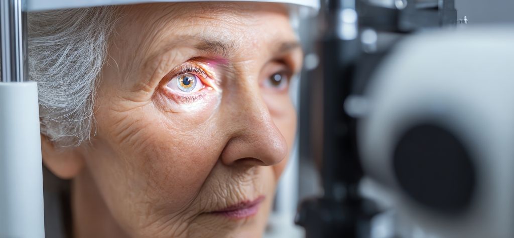
Have you ever looked through a foggy car windshield or a dirty window and struggled to see clearly? That blurred view hints at what living with a cataract is like. Cataracts form when the normally clear lens inside your eye becomes cloudy. Light entering the eye scatters rather than focusing precisely on the retina, leading to dull vision, increased glare and a gradual decline in sight. Cataracts are not a rare condition; more than half of Americans aged 80 or older either have cataracts or have undergone cataract surgery. Worldwide, cataracts remain a leading cause of blindness, especially in low‑income regions where surgical access is limited.
This article dives deep into cataracts—what they are, why they form, who is at risk, how they are treated and, importantly, how to protect your sight. Written in easy‑to‑understand language, it will help you recognize early signs, learn about surgical options and adopt habits to reduce your risk. Keep reading to become empowered to care for your eyes.
What Exactly Is a Cataract?
A cataract is a cloudy area in the eye’s crystalline lens. The lens sits behind your iris and pupil and works like a camera lens, focusing light onto the retina. When we’re young, our lenses are transparent, allowing images to appear sharp. Over time, proteins in the lens can clump together and form opacities. These cloudy patches interfere with the passage of light, creating blurred or hazy vision.
How Cataracts Form
Researchers have discovered several mechanisms behind cataract formation:
- Age‑related changes in lens proteins: After age 40, proteins in the lens may begin to break down or clump together, gradually creating cloudiness.
- Oxidative stress and ultraviolet (UV) exposure: Ultraviolet rays generate free radicals that can damage lens proteins. Long‑term exposure without protection contributes to cataract formation. This is why wearing sunglasses that block 100 % of UVA/UVB rays is so important.
- Metabolic factors: Conditions like diabetes cause elevated blood glucose that leads to sorbitol accumulation in the lens, disrupting its transparency. Tobacco, excessive alcohol and poor nutrition also accelerate oxidative damage.
- Injury or inflammation: Traumatic blows, eye surgery or chronic inflammation can disturb the lens capsule or accelerate protein changes. Radiation treatments can have similar effects.
- Genetic predisposition: Congenital cataracts can be present at birth due to gene mutations, while family history increases risk for age‑related cataracts.
Types of Cataracts
Not all cataracts are identical. Doctors classify them by location and cause:
- Nuclear Sclerotic Cataract – The most common type develops in the center (nucleus) of the lens. It tends to progress slowly and initially may even improve near vision (“second sight”) before eventually causing overall blurring and yellowing of colors.
- Cortical Cataract – Begins as spoke‑like wedges at the edge of the lens cortex and progresses toward the center. People often notice glare around lights and difficulty driving at night.
- Posterior Subcapsular Cataract – Forms at the back of the lens capsule, directly in the path of incoming light, leading to glare and halos around lights. It can progress quickly and is more common in younger people, diabetics and those on long‑term steroid therapy.
- Congenital or Pediatric Cataract – Present at birth or develops during childhood. These can result from genetic mutations, infections in utero or metabolic disorders and may require early surgery to prevent amblyopia (lazy eye).
- Secondary or Traumatic Cataract – Develops after an eye injury, inflammation or due to medications like corticosteroids. They can also occur after eye surgery for glaucoma or other conditions.
Understanding the type helps eye‑care specialists recommend appropriate monitoring and treatment strategies.
Recognizing the Symptoms of Cataracts

Cataracts often develop slowly. Many people do not realize their vision is changing until symptoms become pronounced. The National Eye Institute lists several common signs to watch for:
- Blurry or clouded vision: As the lens clouds, images and text appear fuzzy or hazy. You might feel as if you are constantly looking through dirty glasses.
- Faded or yellowed colors: Colors lose their vibrancy and may appear more brownish or yellow.
- Sensitivity to light and glare: Bright sunlight or headlights produce intense glare, making it uncomfortable to be outdoors or drive at night.
- Halos around lights: Many cataract patients report seeing circles or halos around lamps, headlights or streetlights.
- Difficulty seeing at night: Reduced contrast makes night driving challenging. People often notice they need brighter lights for reading or tasks.
- Double vision in one eye: Cataracts can split incoming light, creating double images in the affected eye.
- Frequent changes in glasses prescription: Rapidly changing or stronger prescriptions are another sign that the lens is becoming less flexible and more opaque.
These symptoms may also accompany other conditions. If you notice any of them, schedule an eye exam. Ophthalmologists can differentiate cataracts from issues like glaucoma, macular degeneration or diabetic retinopathy.
Stat: The Cleveland Clinic notes that nearly 1 in 5 Americans aged 65–74 have cataracts and more than half of those over age 80 have undergone cataract surgery.
Who Is at Risk? — Risk Factors and Causes
Cataracts can affect anyone, but certain factors increase risk. Understanding these helps you take preventive actions.
Age and Demographics
- Age: Cataracts are strongly associated with aging. In the United States, cataract prevalence jumps significantly after age 60.
- Gender: Women may be slightly more likely than men to develop cataracts, especially in older age groups.
- Ethnicity: Research shows higher prevalence in certain populations, including people of African and Hispanic descent.
Medical Conditions and Lifestyle Factors
- Diabetes: Elevated blood glucose causes sugar alcohols to accumulate in the lens. This disrupts clarity and accelerates cataract formation.
- Smoking: Tobacco smoke introduces free radicals that damage lens proteins and reduces levels of antioxidants like vitamin C.
- Alcohol consumption: Excessive alcohol intake contributes to oxidative stress and nutrient deficiencies.
- Obesity and hypertension: These conditions are linked to metabolic changes that impair eye health.
- Nutrition: Diets low in antioxidants (vitamins C and E, carotenoids) and high in refined carbohydrates increase risk.
- Steroid use: Long‑term oral or inhaled corticosteroids can hasten posterior subcapsular cataract development.
- Radiation exposure: Ultraviolet rays, X‑rays and other forms of radiation damage lens proteins.
- Eye injury or surgery: Trauma, inflammation and previous surgeries (e.g., vitrectomy) can lead to secondary cataracts.
- Family history: Genetic predisposition plays a role; congenital cataracts run in families.
Environmental and Socioeconomic Factors
- Sun exposure: People living near the equator or spending significant time outdoors have greater cumulative UV exposure. Failing to wear sunglasses increases risk.
- Low income and limited access to care: Cataracts are the leading cause of blindness in low‑ and middle‑income countries. A global study found that cataract cases surged from 32.8 million in 1990 to 82.2 million in 2021. Age‑specific burdens peaked among people aged 70–74, particularly women.
- Air pollution: Emerging research links particulate matter to increased cataract incidence.
Knowing your risk factors allows you to address modifiable ones and monitor your eye health closely.
The Science Behind Cataract Development

Lens Anatomy and Physiology
The crystalline lens consists of:
- Capsule: A thin, elastic membrane encasing the lens. It holds the lens in place via zonular fibers attached to the ciliary body.
- Cortex: The soft, outer region composed of newly formed lens fibers.
- Nucleus: The dense, central region that accumulates with age.
The lens is avascular (without blood supply). It receives nutrients from surrounding ocular fluids and relies on antioxidants to prevent protein oxidation. Age reduces the lens’s ability to maintain clarity, and clumped proteins scatter light, forming cataracts.
Oxidative Stress and Protein Changes
At the molecular level, oxidative stress is a major driver of cataract formation. Free radicals—unstable molecules generated by UV exposure, smoking, poor diet and inflammation—attack lens proteins and lipids. Damaged proteins lose their solubility and aggregate, forming the characteristic cloudiness of a cataract. Enzymatic defenses like glutathione peroxidase and superoxide dismutase decline with age, making the lens more susceptible to damage.
Role of Nutrition and Vitamins
Research highlights the importance of certain nutrients:
- Vitamin C and E: Powerful antioxidants that neutralize free radicals and maintain lens transparency. Studies show diets rich in fruits (citrus, berries) and vegetables lower cataract risk.
- Carotenoids (lutein and zeaxanthin): These pigments accumulate in the lens and retina, filtering blue light and quenching free radicals. Dark leafy greens, corn and egg yolks are excellent sources.
- Omega‑3 fatty acids: May reduce inflammation and support overall eye health.
- Zinc and selenium: Trace minerals involved in antioxidant enzyme activity.
Diagnosing Cataracts
Early detection is key to preventing vision loss. Eye‑care professionals use several tests to evaluate lens clarity:
- Visual acuity test: Using a standard eye chart to determine how well you see at a distance.
- Slit‑lamp examination: A microscope with a bright light illuminates the front of the eye. It allows the doctor to examine the cornea, iris, lens and anterior chamber.
- Retinal exam: After dilating your pupils, the doctor looks at the retina and optic nerve to rule out other problems such as macular degeneration or diabetic retinopathy.
- Tonometry: Measures intraocular pressure to check for glaucoma.
Regular comprehensive eye exams (every 1–2 years for adults over 60) help catch cataracts and other conditions early.
How Are Cataracts Treated?
When to Consider Surgery
In the early stages, cataracts may cause minimal vision impairment. Updating your eyeglass prescription, using brighter lighting, wearing anti‑glare sunglasses and magnifying lenses can help. When cataracts interfere with reading, driving or daily tasks, surgery becomes the best option. Cataract surgery is the only way to remove a cataract. Your ophthalmologist will recommend surgery when reduced vision affects your quality of life.
Cataract Surgery Explained
During cataract surgery, the cloudy natural lens is removed and replaced with a clear artificial lens called an intraocular lens (IOL). Advances in surgical techniques have made the procedure safe and highly effective.
Phacoemulsification
The most common method, phacoemulsification, uses ultrasound vibrations to break up the cloudy lens. A small incision (2–3 mm) is made, and an ultrasonic probe emulsifies the lens, which is then suctioned out. A folded IOL is inserted through the same incision and unfolds inside the capsular bag. Because the incision is tiny, stitches are rarely needed, and recovery is quick.
- Success rate: Modern extracapsular cataract extraction techniques have improved so much that overall success rates are around 90–95 percent.
- Benefits: Better visual outcomes, minimal discomfort, short surgical time (typically 10–20 minutes), and rapid healing.
Femtosecond Laser‑Assisted Surgery
Newer practices use femtosecond laser technology to perform certain steps of cataract surgery. The laser creates precise incisions, opens the lens capsule and pre‑segments the lens before ultrasound removal. Although this can improve precision and reduce manual skill variations, studies do not show significantly better outcomes compared to standard phacoemulsification. Laser‑assisted surgery is often more expensive and may not be covered by insurance.
Intraocular Lens Options
IOLs come in various designs to match patients’ visual needs:
- Monofocal lenses: Provide clear vision at one distance—usually far. Patients may still need reading glasses.
- Multifocal lenses: Have multiple focal zones to provide distance and near vision. They reduce reliance on glasses but may cause halos or glare.
- Toric lenses: Correct astigmatism and provide distance vision. Additional reading glasses may still be required.
- Accommodating lenses: Flex within the eye to provide a range of focus; they mimic natural lens movement but are more expensive.
Your surgeon will discuss lens options based on your lifestyle and ocular measurements.
Recovery and Healing
Recovery is generally smooth. Many people see improved vision within 24–48 hours. Complete healing takes four to eight weeks. After surgery, you will likely:
- Use prescription eye drops to prevent infection and reduce inflammation.
- Avoid strenuous activities and heavy lifting for a week or two.
- Wear a protective shield while sleeping to avoid rubbing your eye.
- Attend follow‑up visits to monitor healing and check the IOL position.
Risks and Complications

Modern cataract surgery is one of the safest procedures in medicine, but like any surgery, complications can occur. The American Academy of Ophthalmology lists potential risks:
- Eye infection or bleeding
- Swelling of the cornea or retina
- Detached retina (when the retina lifts up from the back of the eye)
- Damage to other parts of the eye
- Persistent pain, blurry vision or seeing halos and dark shadows
- Dislocation of the IOL implant
Most complications are rare and manageable. Serious issues like infection and retinal detachment occur in less than 1 % of cases. Your surgeon will discuss risks and ensure you know when to seek help.
Posterior Capsular Opacification (PCO)
Months or years after surgery, some people develop posterior capsular opacification—sometimes called a secondary cataract. It occurs when the membrane holding the IOL becomes cloudy. Unlike primary cataracts, PCO is easily treated with a quick, painless laser procedure called YAG laser capsulotomy, which restores clear vision.
Living With Cataracts Before Surgery
For many people, cataracts progress slowly. Early lifestyle adjustments can help you continue daily activities:
- Update your prescription: Frequent eye exams will ensure your glasses or contacts provide the best possible vision.
- Use brighter lighting: Add lamps and use daylight bulbs to improve contrast when reading.
- Reduce night driving: Glare from headlights can be dangerous; consider ride‑shares or day travel when possible.
- Utilize magnifying aids: Handheld magnifiers and digital screens with zoom functions enhance reading.
- Protect your eyes: Wear sunglasses that block 100 % UVA/UVB rays and a wide‑brimmed hat when outdoors.
- Manage chronic conditions: Keep blood sugar and blood pressure under control to slow cataract progression.
Preventing Cataracts and Protecting Your Vision
While you cannot stop aging, you can adopt habits that lower your risk of cataracts or delay their progression:
- Wear UV‑blocking sunglasses: Choose glasses that block 99–100 % of UVA and UVB light. This simple step protects the lens from UV‑induced oxidative damage.
- Quit smoking: Tobacco smoke accelerates oxidative damage. Quitting reduces risk and benefits overall health.
- Limit alcohol and maintain a healthy diet: Excess alcohol increases cataract risk. Focus on a diet rich in fruits, vegetables, whole grains, lean proteins and healthy fats.
- Control diabetes and blood pressure: Good glycemic control reduces the sorbitol build‑up that clouds the lens.
- Manage medications: If you take corticosteroids for asthma, arthritis or allergies, consult your doctor about the lowest effective dose. Steroid‑sparing treatments may reduce cataract risk.
- Protect your eyes from injury: Wear protective goggles when playing sports, using power tools or working around chemicals.
- Get regular eye exams: Early detection means you can address small cataracts before they severely impact vision.
The Role of Antioxidants and Lifestyle
Some research suggests that a diet rich in antioxidants may slow cataract development. Vitamin C, vitamin E, lutein and zeaxanthin can be obtained through diet or supplements. However, supplementation should be discussed with an eye‑care professional to ensure safety and proper dosing.
Can Eye Drops Dissolve Cataracts?
There is currently no scientifically proven eye drop that can dissolve or reverse cataracts. Some companies market lanosterol‑based eye drops claiming to clear cataracts, but there is insufficient clinical evidence supporting these claims. At present, surgery remains the only effective treatment.
Cataracts Worldwide: A Public Health Perspective

The burden of cataracts extends beyond individuals; it is a major public health challenge. According to a global analysis, the total number of prevalent cataract cases increased from 32.8 million in 1990 to 82.2 million in 2021. Disability‑adjusted life years (DALYs) attributable to cataracts rose from 2.5 million to 5.2 million. Age‑standardized rates peak in people aged 70–74, and women tend to be more affected than men. Low‑income countries face the highest burden due to lack of surgical access, limited ophthalmologists and inequities in healthcare.
Economic and social impacts include:
- Reduced productivity: Vision loss impairs an individual’s ability to work, travel, cook and perform daily tasks.
- Caregiver burden: Family members may need to assist with tasks or transportation, affecting their work and quality of life.
- Healthcare costs: Untreated cataracts lead to accidents and injuries. However, cataract surgery is cost‑effective and can restore sight, independence and productivity.
International organizations like the World Health Organization (WHO) and Vision 2020 campaign advocate for avoidable blindness programs. Goals include training more eye surgeons, creating community eye health initiatives and providing low‑cost surgeries. Innovations such as manual small‑incision cataract surgery (MSICS) provide effective, affordable procedures in resource‑limited settings.
Research and Future Directions
Advances in Surgical Techniques
- Smaller incision phaco: Surgeons continue refining micro‑incision techniques (< 2 mm) to further reduce healing time and induced astigmatism.
- Robotic and automated surgery: Early studies are exploring robotic assistance for precise surgical steps, potentially improving safety and consistency.
- 3D heads‑up displays: These allow surgeons to operate while viewing a high‑resolution 3D screen instead of looking through a microscope, reducing fatigue and improving posture.
- Adjustable IOLs: Light‑adjustable lenses can be fine‑tuned after implantation using UV light, providing customized vision without additional surgery.
Gene Therapy and Pharmacologic Interventions
Scientists are investigating medications that could delay cataract formation. Research into lanosterol and oxysterol compounds aims to dissolve aggregated lens proteins. Gene therapy targeting proteins involved in maintaining lens transparency is another avenue. While these treatments are not yet ready for clinical use, they offer hope for future non‑surgical options.
Artificial Intelligence for Early Detection
AI algorithms are being developed to detect cataracts and other ocular diseases using smartphone cameras and fundus photography. These tools could help screen populations in remote areas and refer patients for timely care.
Living Well After Cataract Surgery
Many people worry about how life will change after surgery. In reality, cataract removal often leads to brighter colors, sharper vision and renewed independence. Here are tips for post‑surgery success:
- Follow your surgeon’s instructions: Take prescribed eye drops and attend follow‑up visits.
- Protect your eye: Wear sunglasses outdoors and a shield while sleeping in the first week.
- Avoid strenuous activities: No heavy lifting or bending at the waist for several weeks to reduce pressure on the eye.
- Don’t rub your eye: Let the incision heal fully; rubbing can introduce infection or dislodge the IOL.
- Report problems: Call your ophthalmologist if you experience intense pain, increasing redness, decreased vision or flashing lights.
Quality of Life Improvements
Studies consistently show that cataract surgery improves quality of life. People report better mobility, easier reading, renewed ability to drive and participate in hobbies like sewing and golfing. Many patients also require fewer hours of caregiver support. Because cataract surgery corrects refractive errors, some individuals find that they no longer need thick glasses and enjoy clear, unaided vision.
Conclusion: Protecting Your Vision for Life
Cataracts are a natural part of aging, but they are not inevitable. By understanding the risk factors—age, UV exposure, diabetes, smoking and more—you can make informed choices that protect your eyes. Recognize symptoms early; don’t ignore blurry vision, glare or frequent prescription changes. Regular eye exams are crucial because cataracts are highly treatable. Modern surgery is safe and effective, boasting success rates above 90 percentpmc.ncbi.nlm.nih.gov. Advances in IOL technology mean you may enjoy better vision than before cataracts developed.
Most importantly, adopt preventive habits today: wear UV‑blocking sunglasses, eat antioxidant‑rich foods, avoid smoking and control systemic diseases. Spread awareness within your community—especially among older relatives—so that cataracts do not steal anyone’s sight unnecessarily. Remember, clear vision enhances your quality of life, independence and connection to the world. Take care of your eyes, and they will continue to serve you well for decades.