
A New Frontier for Eye Health
Imagine a world where your phone or your local clinic can alert you to subtle changes in your eyes long before you notice blurriness or blind spots. That is not science fiction; it is the promise of artificial intelligence (AI) in eye care. Using machine‑learning algorithms and high‑resolution imaging, researchers are training computers to spot the tiniest signs of disease and even predict who may lose vision in the future. This ability could transform how we prevent blindness and manage conditions like diabetic retinopathy, age‑related macular degeneration and glaucoma.
In this article, written in a friendly tone for readers of all ages, we will explore whether AI can indeed forecast vision loss before you see any symptoms. We will discuss why early detection matters, how AI models work and where the technology currently stands. We will also consider potential benefits, challenges, ethical issues and practical tips for protecting your eyes. Our goal is to help you understand how cutting‑edge technologies could shape the future of eye health.
Why Early Detection of Eye Diseases Matters
Vision loss affects millions of people. According to the Centers for Disease Control and Prevention (CDC), approximately 7 million people in the United States have vision impairment, including 1 million with blindness. Another 4.2 million Americans aged 40 or older have uncorrectable vision impairment, a number projected to more than double by 2050. Nearly 93 million adults in the U.S. are at high risk for serious vision loss, but only half visited an eye doctor in the past year. The economic cost of major vision problems is expected to reach $373 billion by 2050.
Eye diseases rarely cause pain in early stages, making them easy to ignore. Conditions like diabetic retinopathy, age‑related macular degeneration (AMD) and glaucoma can progress silently until irreversible damage occurs. Early detection and timely treatment can prevent or delay vision loss. For example, 90 % of blindness in U.S. adults caused by diabetes is preventable. Regular comprehensive eye exams and proper disease management are essential, but many people face barriers such as cost, lack of awareness or limited access to specialists.
AI technologies offer a way to bridge these gaps by making screening faster, cheaper and more accurate. Before we delve into the AI, let’s briefly review some common eye conditions that can lead to vision loss.
Key Eye Conditions Leading to Vision Loss
- Diabetic retinopathy (DR) – Damages blood vessels in the retina due to high blood sugar. Early stages have no symptoms, but advanced DR can cause blurred vision, floaters and even blindness.
- Age‑related macular degeneration (AMD) – Deteriorates the central part of the retina (the macula), leading to loss of sharp central vision. It is a leading cause of blindness among older adults.
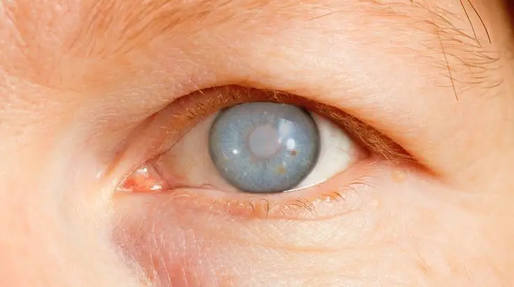
- Glaucoma – A group of diseases that damage the optic nerve, often because of increased pressure inside the eye. Glaucoma causes gradual loss of peripheral vision and is often called the “silent thief” because symptoms appear late.
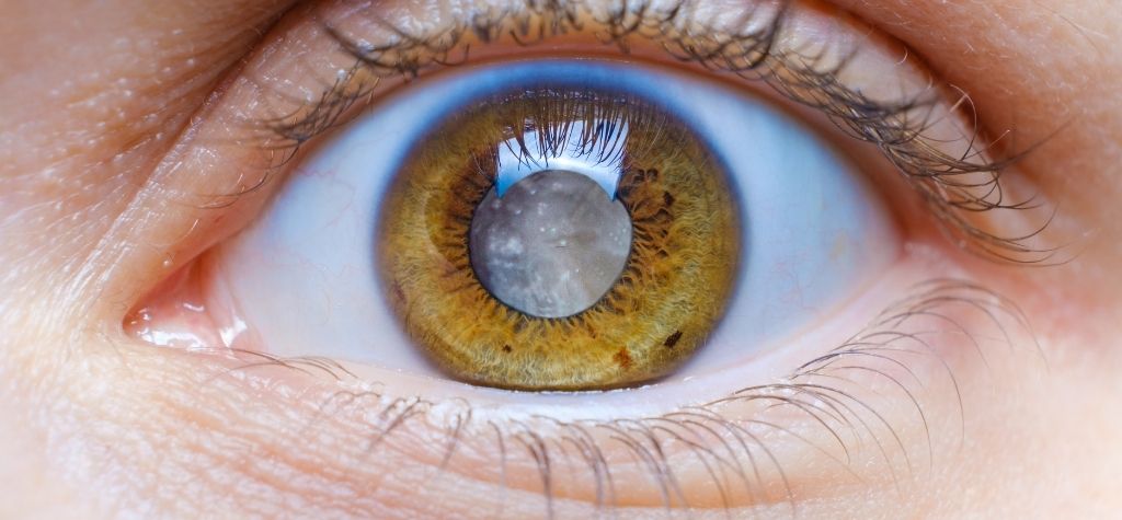
- Keratoconus – A condition in which the cornea bulges outward, causing blurred and distorted vision. It typically appears in teenagers and young adults and can lead to severe vision loss if untreated.
How Artificial Intelligence Works in Eye Care

The Basics of AI and Deep Learning
Artificial intelligence refers to computer systems capable of performing tasks that normally require human intelligence, such as recognizing patterns or making predictions. In eye care, the most common type of AI is deep learning, a form of machine learning that uses convolutional neural networks (CNNs) to analyze images. These networks consist of layers that process raw pixel data from retinal photographs or optical coherence tomography (OCT) scans, extract features and classify patterns associated with disease.
Training a deep‑learning model requires thousands or even millions of labeled images. The model learns which combinations of pixel patterns correspond to healthy eyes and which indicate disease. Once trained, the AI can analyze new images quickly and suggest whether a person has signs of a disease, how severe it is and sometimes how likely it is to progress.
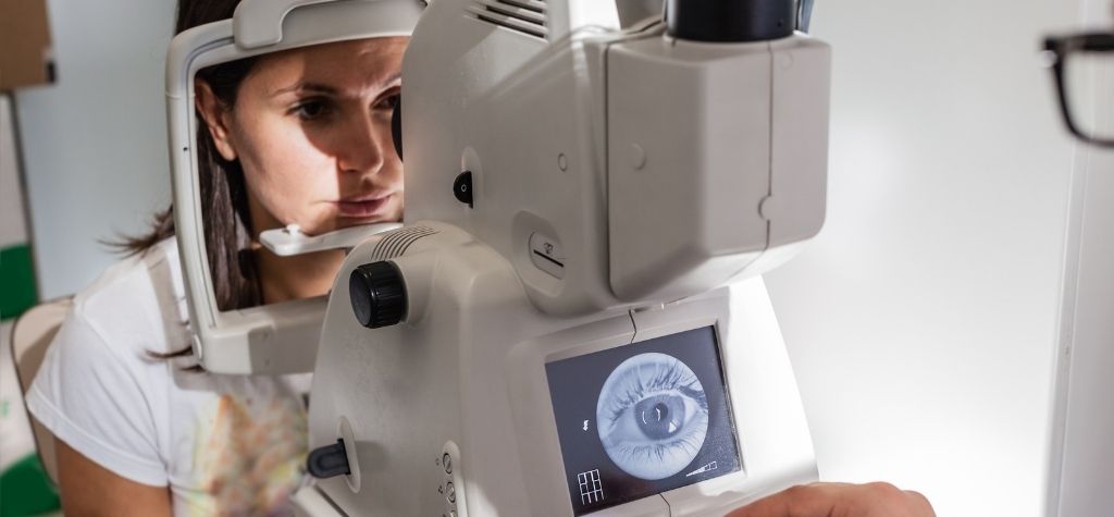
Data Sources: Fundus Photographs and OCT
Two imaging modalities dominate AI applications in ophthalmology:
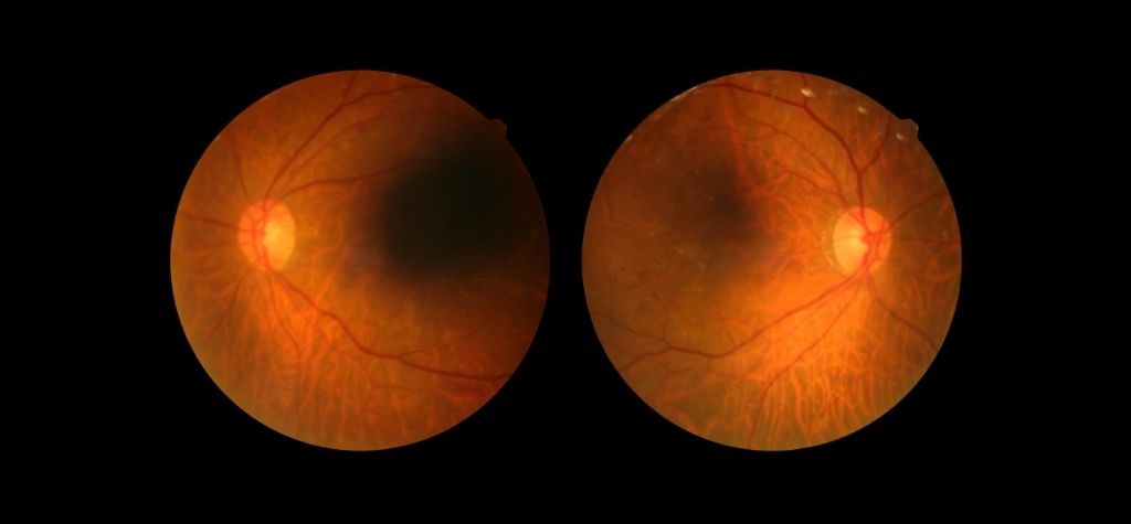
- Color Fundus Photography (CFP) – A wide‑angle photograph of the retina captured through a dilated pupil. Fundus photos highlight blood vessels, the optic disc and the macula and are widely used to screen for diabetic retinopathy and AMD.
- Optical Coherence Tomography (OCT) – Uses light waves to create cross‑sectional images of the retina. OCT provides three‑dimensional information about retinal layers and is essential for diagnosing and monitoring diseases like AMD and glaucoma.
AI models can analyze these images at scale. For example, the National Eye Institute (NEI) developed a novel AI‑based method called parallel discriminator generative adversarial network (P‑GAN). When trained on thousands of adaptive optics OCT images, P‑GAN reduced imaging acquisition and processing time by about 100‑fold and increased image contrast 3.5‑fold. Such improvements make it easier to capture high‑quality images quickly, enabling earlier detection of subtle retinal changes.
From Detection to Prediction
Early AI systems focused on diagnosing disease—determining if someone has diabetic retinopathy or AMD from retinal images. More recent models aim to predict future outcomes. For example, AI algorithms can analyze the current state of the retina and forecast whether a person is likely to develop late‑stage AMD or whether their glaucoma will worsen. These predictive models often combine imaging with demographic data (age, sex) and clinical information (blood pressure, blood glucose) to generate risk scores.
Predictive AI holds tremendous promise because it allows clinicians to intervene before patients experience symptoms or significant vision loss. Let’s explore some specific conditions where AI is making headway.
AI for Diabetic Retinopathy: Screening at Scale
Why DR Screening Matters
Diabetic retinopathy is one of the most common causes of blindness among working‑age adults. Early stages are asymptomatic, but timely treatment (such as laser therapy or injections) can prevent or delay vision loss. Regular screening is essential, yet many patients with diabetes skip annual eye exams. AI can help fill this gap by enabling automated screening in primary care offices and community settings.
FDA‑Approved AI Screening Devices
In 2018 the U.S. Food and Drug Administration (FDA) cleared IDx‑DR, the first autonomous AI device for detecting more than mild diabetic retinopathy. According to the De Novo classification summary, IDx‑DR is indicated for use by health care providers to automatically detect more than mild diabetic retinopathy in adults with diabetes who have not previously been diagnosed with the disease. It works with a specific fundus camera (Topcon NW400) and analyzes retinal images. If the algorithm detects disease, the patient is referred to an eye care provider; if no disease is detected, the patient is advised to recheck later.
Another FDA‑cleared device is EyeArt (from Eyenuk Inc.), which screens for diabetic retinopathy and diabetic macular edema using fundus photographs. A large study described by Riedl and colleagues noted that EyeArt reports 96 % sensitivity and 88 % specificity for detecting diabetic retinopathy and macular edema, and 97 % sensitivity for detecting vision‑threatening disease. These devices can operate without an eye specialist on-site, making screening accessible in primary care clinics and pharmacies.
AI Screening Accuracy vs. Human Experts
Do AI devices match the performance of human graders? A cohort study cited in the review by Riedl et al. compared AI algorithms to human graders in identifying glaucoma from fundus images. The AI correctly identified glaucoma in 88–90 % of cases versus 79–81 % by human graders. For diabetic retinopathy, the IDx‑DR system demonstrated a diagnostic sensitivity of 87.2 % and specificity of 90.7 %, while EyeArt achieved 96 % sensitivity and 88 % specificity. These results indicate that AI can match or surpass human performance, at least for specific tasks.
Benefits Beyond the Clinic
Automated screening may reduce missed cases by bringing retinal exams closer to patients. It can ease the disease burden by allowing automated CFP‑based screening programs in primary care offices. Such programs are especially valuable in underserved areas where access to ophthalmologists is limited. Patients with diabetes could have their eyes photographed during routine doctor visits, with AI providing an immediate risk assessment.
However, AI screening is not perfect. The FDA summary notes that IDx‑DR does not detect other eye diseases and should not replace a comprehensive eye exam. Patients still need regular visits to an eye care provider, especially because people with diabetes are also at increased risk of glaucoma and cataract. AI helps triage patients but does not eliminate the need for human oversight.
AI for Age‑Related Macular Degeneration: From Diagnosis to Risk Prediction
Deep Learning Models for AMD
Age‑related macular degeneration (AMD) is a leading cause of vision loss among older adults. There are two forms—dry (atrophic) and wet (neovascular). Treatments exist to slow progression, but they are most effective when started early. Researchers in the NIH Intramural Research Program (IRP) have developed deep‑learning algorithms that diagnose AMD and classify its severity using color photographs of the retina with greater accuracy than retina specialists. The same algorithms detect important disease features that would otherwise require advanced imaging techniques.
Predicting Who Will Progress
Beyond diagnosis, the IRP team created models that use a patient’s data to predict their risk of progressing to advanced AMD over the next decade. These algorithms incorporate information from images, genetic data, demographics and clinical variables. The models allow clinicians to identify high‑risk patients and schedule closer monitoring or early treatment. As the IRP report notes, AI algorithms can match or exceed specialist evaluations, providing earlier diagnoses and enabling improved clinical outcomes.
Making the Models Accessible
A key advantage of the IRP effort is that the researchers made their algorithms publicly available and embedded them in desktop software for research use. Open access encourages other institutions and companies to test and refine the models, potentially speeding translation into clinical practice. Yet there are still regulatory hurdles; as of early 2026, there is no FDA‑approved autonomous AI device for AMD screening. In practice, AI tools assist ophthalmologists rather than replace them.
Can AI Detect the First Signs of AMD?
Researchers are exploring whether AI can identify subclinical changes that precede visible lesions. In one example, the RETFound foundation model described by Riedl et al. uses self‑supervised learning on unlabeled retinal images to detect early signs of common retinal diseases. Meanwhile, at the National Eye Institute, the P‑GAN technique discussed earlier speeds up imaging of the retinal pigment epithelium (RPE)—tissue that supports photoreceptors. Because diseases like AMD often begin with RPE changes, AI‑enhanced imaging could reveal pathology earlier than conventional methods.
AI for Keratoconus: Predicting Who Needs Treatment
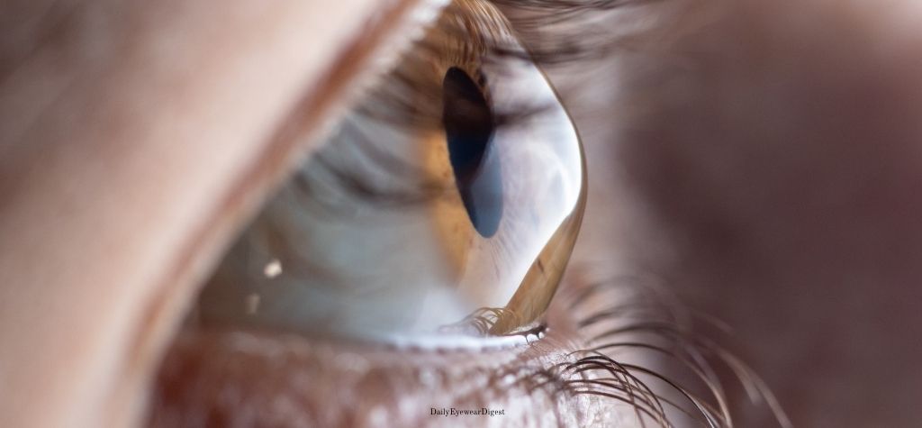
Keratoconus is a progressive thinning and bulging of the cornea. It usually develops in teenagers and young adults and can lead to severe vision loss or the need for a corneal transplant. Treatment options include rigid contact lenses, corneal cross‑linking (which strengthens corneal fibers) and corneal transplantation. Knowing which patients will progress quickly helps doctors decide whether to start treatment early.
Research from Moorfields Eye Hospital and UCL
At the European Society of Cataract and Refractive Surgeons (ESCRS) 2025 conference, researchers from Moorfields Eye Hospital and University College London (UCL) presented a study where they trained AI on 36,673 OCT images from 6,684 patients. The algorithm accurately predicted whether a keratoconus patient’s condition would deteriorate or remain stable using data from the first visit alone. With information from a second visit, it correctly categorized up to 90 % of patients into high‑risk and low‑risk groups.
The study’s lead author, Dr. Shafi Balal, explained that currently, doctors cannot easily distinguish which patients will progress and require cross‑linking and which can be monitored. As a result, patients undergo frequent monitoring and often receive treatment after vision has already worsened. The AI model may change that by enabling preventive cross‑linking before irreversible damage occurs, while sparing low‑risk patients from unnecessary procedures. The technology still needs further safety testing, but it marks a major step toward personalized care for keratoconus.
Implications for Young Patients
Keratoconus affects mostly young, working‑age individuals. Early intervention can preserve vision and avoid the need for corneal transplants, which carry surgical risks and long recovery times. AI tools could enable eye clinics or even mobile screening units to identify patients who need cross‑linking early, improving quality of life and reducing healthcare costs.
AI for Glaucoma: Identifying High‑Risk Patients and Tracking Progression
The Challenge of Predicting Glaucoma
Glaucoma is a major cause of irreversible blindness worldwide. It involves progressive damage to the optic nerve, often due to elevated intraocular pressure. The disease can progress slowly, and visual field tests may take years to detect moderate changes. Clinicians need better ways to identify which patients are at risk of rapid progression so they can tailor treatment accordingly.
Hyperspectral Imaging and AI for Biomarkers
Researchers at the Centre for Eye Research Australia (CERA) are investigating advanced imaging and AI to find biomarkers of sick retinal cells. Associate Professor Zhichao Wu and colleagues developed a hyperspectral camera that captures how retinal cells reflect light. Their goal is to detect cells that are beginning to die before significant damage occurs. In the first phase of their study, they use hyperspectral imaging and AI to identify biomarkers that predict glaucoma progression.
Compressing Detection from Six Years to Six Months
Currently, doctors rely on visual field tests every six months, which can take six years to detect moderate rates of progression. Wu’s team aims to compress this timeframe from six years to six months by using widefield OCT imaging combined with AI. The AI produces a heatmap of sick cells to show where damage is likely to occur and how close it is to the central vision. Such information could help ophthalmologists prioritize treatment for patients most at risk and monitor therapy effectiveness.
Future Prospects for Glaucoma AI
The CERA researchers emphasize that they are still in the early stages and that there are currently no known biomarkers that can accurately predict glaucoma progression. The study is ongoing and will follow participants for two years. Nonetheless, the goal of turning a lengthy monitoring process into a short, actionable prediction demonstrates the potential of AI for glaucoma care.
AI and Retinal Imaging for Systemic Health Prediction
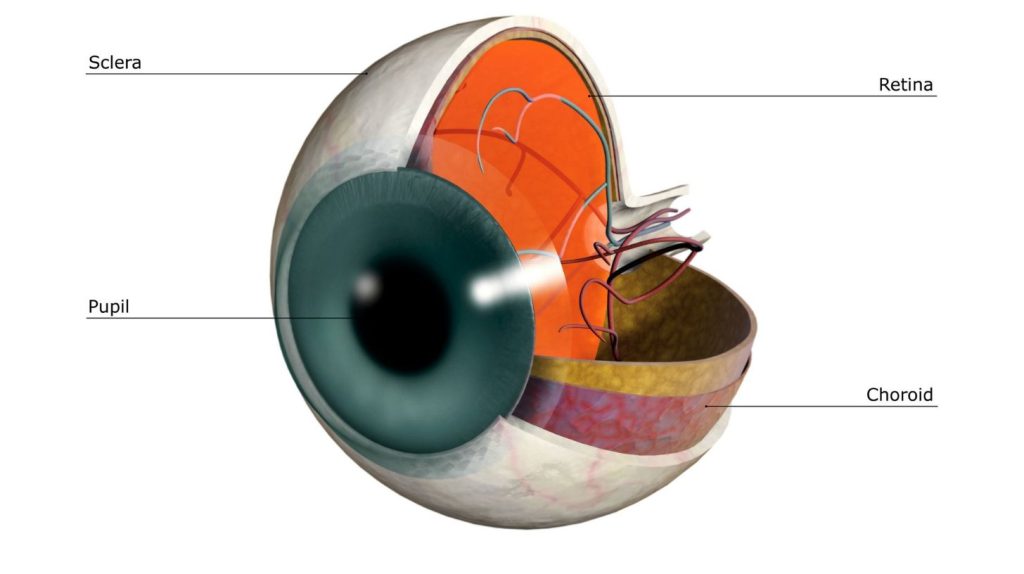
The Retina as a Window to the Body
Ophthalmologists often say that the eye is the only place where doctors can directly see blood vessels without an invasive procedure. Changes in retinal vasculature can reflect systemic conditions like hypertension, diabetes and even neurodegenerative diseases. AI researchers are leveraging this connection to develop oculomics—the use of retinal imaging to infer broader health indicators.
Predicting Cardiovascular and Metabolic Risk
In the review by Riedl and colleagues, researchers noted that AI analysis of color fundus photographs showed correlations between retinal vascular changes and cardiovascular health. They suggested that automated tools could screen populations for heart disease by recognizing patterns of retinal microvasculature. An AI model even predicted age, systolic blood pressure, body mass index and HbA1c levels from retinal images. Another model was trained to predict major cardiovascular events with an area under the curve (AUC) of 0.70.
Early Detection of Neurodegenerative Diseases
Researchers are also exploring retinal biomarkers for Alzheimer’s disease, Parkinson’s disease and multiple sclerosis. For example, vascular density measured by OCT‑angiography (OCT‑A) was significantly reduced in patients with Alzheimer’s disease compared to controls. Studies have achieved detection of subclinical Alzheimer’s disease with OCT‑A in small cohorts, suggesting that the retina might reveal neurodegeneration before cognitive symptoms appear. In Parkinson’s disease and multiple sclerosis, specific OCT‑A parameters show promise as early biomarkers.
Limitations of Oculomics
While the prospect of predicting heart attacks or neurological disorders from eye scans is exciting, it remains an area of active research. The AUC values (e.g., 0.70) indicate moderate predictive power, and the models must be validated in diverse populations. Using retinal images to infer systemic health also raises ethical concerns about privacy and data use, which we will discuss later.
Home Monitoring and Telemedicine: AI on Your Smartphone
Portable Devices and Home OCT
Advances in miniaturization and AI have enabled the development of portable OCT devices with built‑in AI algorithms. Riedl et al. note that patient‑operated OCT devices with automated image evaluation have become available for home monitoring. When these devices detect significant changes, they can prompt patients to consult their ophthalmologist. This approach is especially useful for chronic conditions like AMD, where monthly injections or treatments are needed, and for patients who live far from specialists.
Smartphone‑Based Eye Exams
Several companies and researchers are working on smartphone attachments that allow users to take fundus photographs or measure vision. AI algorithms analyze these images and provide preliminary assessments. While smartphone exams cannot replace a comprehensive eye exam, they may encourage more people to monitor their eye health and seek care earlier.
Telemedicine and Access to Care
Telemedicine platforms combined with AI can link patients in remote or underserved areas to specialists. For example, images taken by local healthcare workers can be uploaded to a cloud service where AI assists in triaging cases. Only patients who need further evaluation are referred, reducing travel costs and wait times. This model could improve eye care equity, especially in regions where ophthalmologists are scarce.
Potential Benefits of AI in Predicting Vision Loss
AI has the potential to transform eye care in several ways:
- Earlier diagnosis and treatment: By detecting disease or risk earlier, AI may enable interventions that prevent or delay vision loss.
- Increased access: Automated screening and home monitoring can reach people who do not have easy access to specialists.
- Improved efficiency: AI can handle large volumes of images quickly, freeing up clinicians to focus on complex cases.
- Personalized care: Predictive models can identify high‑risk individuals and tailor monitoring schedules and treatment plans.
- Systemic health insights: Oculomics research may allow eye scans to serve as a noninvasive screening tool for cardiovascular or neurological diseases.
Challenges and Limitations
While the benefits are substantial, there are important challenges to consider:
- Data Quality and Diversity: AI models require large, high‑quality datasets. If the data come from a narrow patient demographic, the models may not generalize well to other populations. Riedl et al. highlight the lack of diversity in algorithmic training as a challenge.
- Regulatory and Legal Issues: The FDA has only approved a handful of AI devices. Developers must demonstrate safety and effectiveness, handle data privacy and ensure that models do not perpetuate bias.
- Integration into Clinical Workflows: Clinicians need training to interpret AI outputs and integrate them into decision‑making. Overreliance on AI could lead to missed diagnoses or delayed treatment.
- Ethical Concerns: Using retinal images to predict systemic conditions raises questions about consent and privacy. Patients should know how their data will be used and stored.
- Cost and Access: Although AI screening can reduce costs in the long term, initial deployment of devices and training may be expensive. Ensuring that AI benefits reach underserved communities requires investment.
How to Protect Your Vision Today

While AI technologies are advancing, there are steps you can take now to protect your vision:
- Get Regular Eye Exams: Adults over 60, people with diabetes, and those with a family history of eye disease should have comprehensive eye exams at least once a year. Early detection remains the best defense against vision loss.
- Manage Chronic Conditions: Keep blood sugar and blood pressure under control to reduce the risk of diabetic retinopathy and hypertensive retinopathy. Follow your doctor’s recommendations for managing cholesterol and other health parameters.
- Wear UV‑Blocking Sunglasses: The National Eye Institute advises wearing sunglasses that block 99–100 % of UVA and UVB rays to protect your eyes from ultraviolet light. The CDC notes that UV exposure increases the risk of blinding eye diseases and recommends wraparound sunglasses that block both UVA and UVB.
- Quit Smoking: Smoking increases the risk of AMD and cataract. If you smoke, talk to your healthcare provider about quitting strategies.
- Maintain a Healthy Lifestyle: Eat a balanced diet rich in leafy greens, fish and colorful fruits; exercise regularly; and maintain a healthy weight. These habits support overall health and may reduce the risk of eye disease.
- Learn About AI Tools: Ask your eye doctor whether AI screening or home monitoring devices are available in your area. Be informed about how your data will be used and stored.
Ethical and Social Implications of AI in Eye Care
Privacy and Data Security
Retinal images are biometric data, meaning they are unique to you. When used for AI models, these images may reveal not only eye diseases but also systemic health conditions. Healthcare providers and AI developers must protect patient privacy by using secure data storage, encryption and anonymization. Regulations like the Health Insurance Portability and Accountability Act (HIPAA) in the U.S. set standards for data handling, but new guidelines may be needed as AI capabilities expand.
Bias and Fairness
If AI algorithms are trained on data from predominantly light‑skinned or certain ethnic populations, they may perform poorly on other groups. This could worsen health disparities. Researchers must ensure that training datasets are diverse and include enough examples from different ages, ethnicities and disease stages. Regulators should require developers to report model performance across subgroups.
The Role of Clinicians
AI is a tool to assist, not replace, human expertise. Ophthalmologists bring years of training, clinical judgment and the ability to consider patient preferences. While AI can provide risk scores or suggest diagnoses, clinicians must interpret the results within the context of each patient’s history. Shared decision‑making—where doctors and patients discuss the pros and cons of treatment options—remains essential.
Access and Equity
AI can increase access to care, but only if devices and training are distributed fairly. There is a risk that wealthy regions will adopt AI quickly while under‑resourced areas lag behind. Governments, health systems and nonprofits should invest in technology deployment, staff training and public awareness to ensure equitable benefits.
The Future of AI in Vision Care
Integration of Multimodal Data
Future AI models will likely integrate data from multiple sources, including fundus photography, OCT, genetic tests, lifestyle factors and electronic health records. Combining these datasets could improve predictive accuracy and provide personalized risk profiles. For example, an AI system might consider a patient’s retinal images, age, blood sugar levels and genetic risk factors to forecast vision loss more precisely than any single test.
Real‑Time Decision Support
As AI models become more sophisticated, they could provide real‑time decision support during eye exams or surgery. For instance, AI could analyze an OCT scan as the patient sits in the exam chair and immediately display a risk score and recommended follow‑up schedule. In surgery, AI‑guided instruments could improve precision and reduce errors【784569523403419†L149-L126】, as referenced by some ophthalmology surveys.
Home Monitoring and Patient Engagement
Home OCT devices and smartphone apps will continue to evolve. Some may offer guided image capture, automated analysis and direct communication with healthcare providers. Such tools could empower patients to take an active role in monitoring their eye health.
Broadening the Scope: From Eyes to Systemic Health
The concept of oculomics suggests that retinal imaging may become a routine part of general health screening. Imagine a primary care clinic where a quick eye scan not only assesses your risk for vision loss but also screens for hypertension, diabetes or dementia. AI models are already showing moderate success in predicting blood pressure and major cardiovascular events. As algorithms improve and are validated, they could transform preventive medicine.
Conclusion: Can AI Predict Vision Loss Before You Notice It?
So, can artificial intelligence really predict vision loss before you notice it? The answer is yes—in certain situations, with important caveats. AI algorithms already match or surpass human experts at detecting and grading diabetic retinopathy, and they are beginning to forecast who will develop severe forms of AMD or keratoconus. Emerging research shows that AI can compress glaucoma monitoring timelines and identify retinal changes that signal systemic diseases.
However, the technology is still evolving. Models must be trained on diverse datasets, validated across populations and integrated thoughtfully into clinical practice. AI should complement, not replace, regular eye exams and professional judgment. Moreover, ethical considerations—privacy, fairness and access—must guide the deployment of AI in eye care.
As a patient, you can look forward to a future where an eye scan may reveal not only how your eyes are doing but also offer a window into your overall health. Until then, take care of your eyes through regular exams, a healthy lifestyle and awareness of new tools that may help your doctor keep you seeing clearly for years to come.